Predicting mosquito infection from Plasmodium falciparum gametocyte density and estimating the reservoir of infection
- Imperial College London, United Kingdom
- Radboud University Nijmegen Medical Centre, The Netherlands
- London School of Hygiene and Tropical Medicine, United Kingdom
- University of Edinburgh, United Kingdom
- Centre National de Recherche et de Formation sur le Paludisme, Burkina Faso
Figures
Figure 1
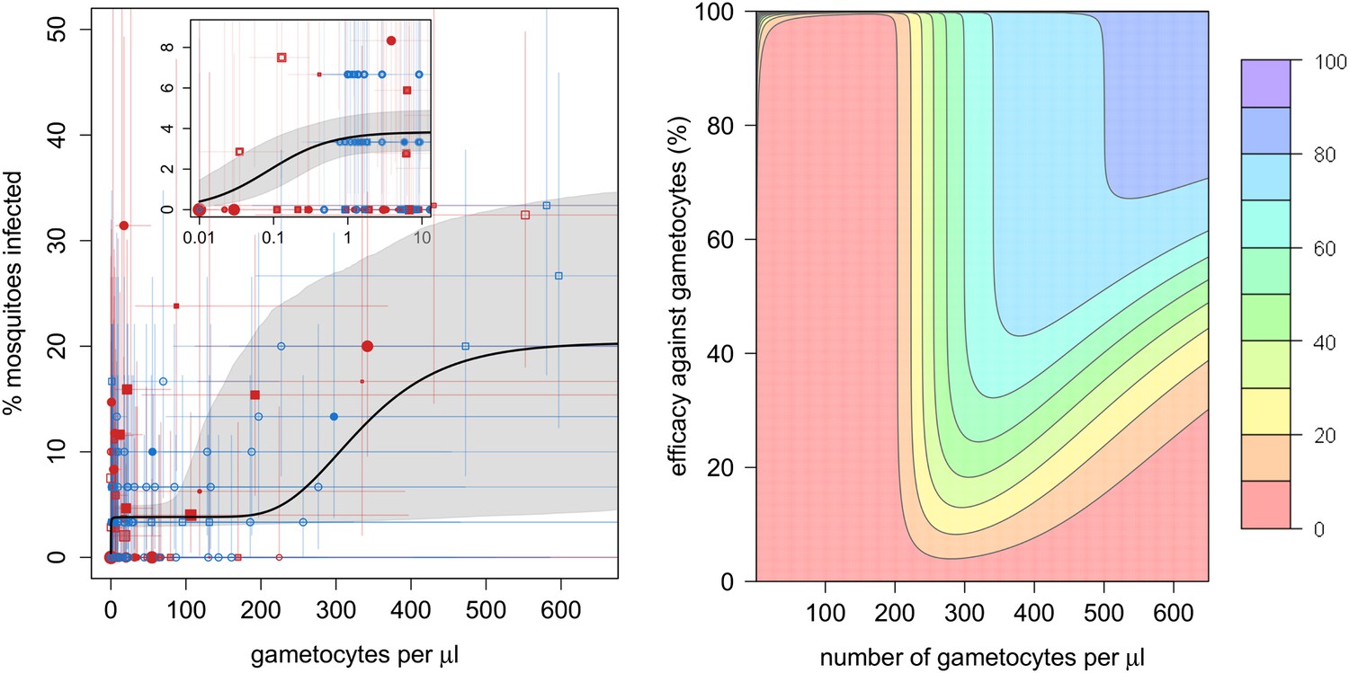
The relationship between gametocyte density and mosquito infection and the impact this will have on the effectiveness of gametocytocidal interventions.
(A) The relationship between Plasmodium falciparum gametocyte density and the percentage of Anopheles gambiae mosquitoes that develop oocysts. Point colour, shading, and shape denote characteristics of the blood donor, such as location (blue = Burkina Faso; red = Kenya), asexual parasite density as measured by microscopy (no fill colour = none detectable, light shading = 1–1000 parasites per microlitre, dark shading ≥1000 µl−1), or host age (<6 years old = square, ≥6 years old = circle). The size of the point is proportional to the number of mosquitoes dissected. Coloured horizontal and vertical lines indicate 95% Bayesian credible intervals (CIs) around point estimates. The solid black line indicates the best-fit model, whereas the grey shaded area indicates the uncertainty around this line. The inset shows the relationship at very low gametocyte densities (on a logarithmic scale). The outputs show the shape of the relationship for a child with no detectable asexual parasites. A full description of data used to fit the model are given in Figure 1—source data 1. Regression coefficients and a measure of goodness-of-fit of the different models are given in Figure 1—source data 2. Panel (B) shows how efficacious transmission-reducing interventions would need to be (on the vertical axis) at decreasing gametocyte density in order to reduce human to mosquito transmission (contour lines). The best-fit line from (A) is used to illustrate the percentage reduction in mosquito infection that would be achieved according to the pre-intervention host’s gametocyte density (on the horizontal axis) for an intervention, which reduces gametocyte density by a given percentage (which is assumed to be constant over different gametocyte densities). The colours represent the percentage reduction in mosquito infection that would be achieved, ranging from red (low, 0–10% reduction) to darker hues of blue (high, 70–80% and 80–90% reduction, see legend). The 90–100% reduction in transmission is hardly visible and would correspond to nearly 100% efficacious interventions at the top of the graph.
-
Figure 1—source data 1
Description of the direct feeding assay data used to estimate mosquito infection.
- https://doi.org/10.7554/eLife.00626.004
-
Figure 1—source data 2
Best-fit model and parameters.
Lower DIC values indicate a more parsimonious fit to the data. All models were fitted with gametocyte density on the arithmetic scale. There was no evidence of any difference between study sites (DIC of best-fit model allowing mosquito infection to vary between study location = 1042). The best-fit model on the logarithmic scale was the Gompertz model (DIC = 1053).
- https://doi.org/10.7554/eLife.00626.005
Figure 2
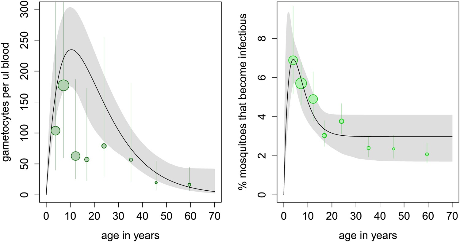
Age patterns of gametocyte density and estimates of the age profile of the human reservoir of infection.
(A) Results from a cross-sectional survey conducted in a high transmission setting in Burkina Faso showing how the mean number of gametocytes per microlitre of blood (including 0s) changes with host age. The distribution of gametocytes among hosts is highly overdispersed. A full description of data used to fit the model are given in Figure 2—source data 1 with the best-fit parameter estimates shown in Figure 2—source data 2. The relationship between gametocyte density and oocyst prevalence shown in Figure 1A is used to predict the percentage of mosquitoes that will become infected after biting a host of a certain age. This is shown in panel (B), which can be interpreted as the contribution of each age group towards the human to mosquito transmission. In both figures, the black solid line shows the best-fit model and the grey shaded area indicates the uncertainty (95% Bayesian credible Interval, CI) having fitted the model to the individual data (a total of 412 individuals). For illustrative purposes, the data are grouped into seven bins, namely 0–5, 5–10, 10–15, 15–20, 20–30, 30–40, 40–50, and ≥50 year olds, and the size of the point is proportional to the number of individuals in the group. In Figure 1A, the 10–15 and 15–20 year groups appear lower than the best-fit line due to sampling artefacts generated by the highly overdispersed data. Vertical lines indicate the 95% CI around grouped estimates.
-
Figure 2—source data 1
Description of the cross-sectional survey on 412 hosts carried out in Burkina Faso.
Asexual parasite density was estimated by microscopy while gametocyte density was estimated using QT-NASBA.
- https://doi.org/10.7554/eLife.00626.007
-
Figure 2—source data 2
Parameter estimates for the age profile of gametocyte density and the force of infection.
- https://doi.org/10.7554/eLife.00626.008
Tables
Table 1
Notation of statistical and mathematical models
| Notation | Description | Equation |
| Y | Time to positivity (TTP) readout generated by QT-NASBA | Equation 1 |
| x | Known density of gametocytes per millilitre (generated using dilution series) | Equation 1 |
| Estimate of (unknown) gametocyte density on the logarithmic scale | Equation 2 | |
| Estimate of gametocyte density on the arithmetic scale | Equation 2 | |
| β0 | Intercept of the calibration line fitted to the dilution series | Equation 1 |
| β1 | Gradient of the calibration line fitted to the dilution series | Equation 1 |
| σ2 | Intra-assay variance measuring the accuracy with which the calibration line fits the TTP estimates from the dilution series | Equation 1 |
| g | Proportion of mosquitoes developing oocysts | Equation 3 |
| Saturating function determining the shape of the initial relationship between gametocytes and the proportion of mosquitoes developing oocysts | Equation 4 | |
| Function determining the shape of the relationship between gametocytes and proportion of mosquitoes developing oocysts. Subscript i indicates the functional form used, be it , where constraining different parameters can generate a range of different shapes (constant α1 = 0, linear α2 = 1; α3 = 0, power α3 = 0, hyperbolic α1 > 0 α2 = 1 α3 > 0, or sigmoid α2 > 1) or , which generates a Gompertz (sigmoid-like) function | Equation 3 | |
| μ | Vector of regression coefficients | Equation 3 |
| z | Vector of dummy variables denoting donor blood characteristics, z1 = asexual parasite density (0 = undetected, 1 = low, 2 = high), z2 = host age (0 = younger than 6 years old, 1 = 6 or older), z3 = study locale (0 = Burkina Faso, 1 = Kenya) | Equation 3 |
| h(A) | Function describing how gametocyte density and the reservoir of infection change with host age (A). Shape determined by parameters τ, ψ, and ω | Equation 5 |
Download links
A two-part list of links to download the article, or parts of the article, in various formats.
Downloads (link to download the article as PDF)
Open citations (links to open the citations from this article in various online reference manager services)
Cite this article (links to download the citations from this article in formats compatible with various reference manager tools)
Predicting mosquito infection from Plasmodium falciparum gametocyte density and estimating the reservoir of infection
eLife 2:e00626.
https://doi.org/10.7554/eLife.00626


