Phosphorylation of β-arrestin2 at Thr383 by MEK underlies β-arrestin-dependent activation of Erk1/2 by GPCRs
- CNRS, UMR-5203, Institut de Génomique Fonctionnelle, France
- INSERM, U1191, France
- Université de Montpellier, France
- INRA, UMR85, Unité Physiologie de la Reproduction et des Comportements, France
- CNRS, UMR7247, France
- Université François Rabelais, France
Figures
Figure 1 with 5 supplements
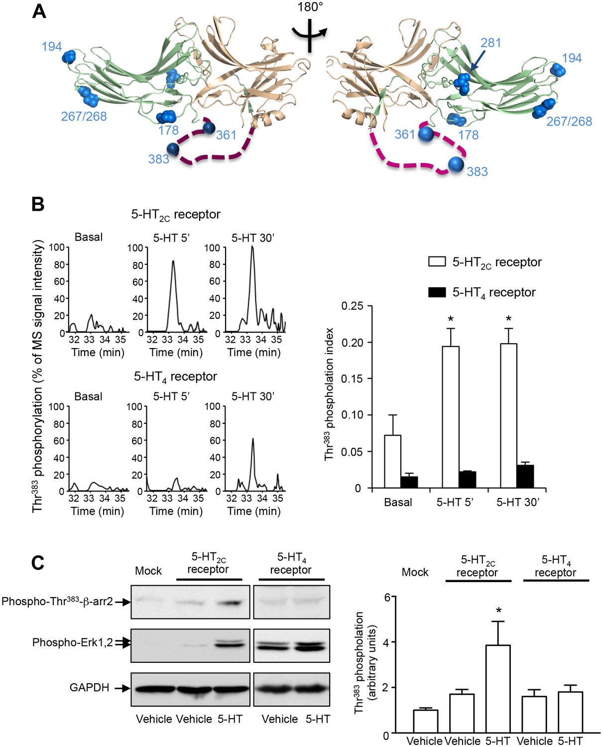
5-HT2C and 5-HT4 receptor stimulation promotes β-arrestin2 phosphorylation in HEK-293 cells.
(A) Ribbon diagram of rat β-arrestin2 showing the position of phosphorylated residues identified by LS-MS/MS. (B) Representative extracted ion chromatograms of the EIDIPVDTNLIEFDTNYAp383TDDDIVFEDFAR peptide from YFP-tagged β-arrestin2 in cells expressing 5-HT2C or 5-HT4 receptor and challenged with vehicle (Basal) or 5-HT (1 and 10 µM, respectively) for 5 or 30 min. Two other independent experiments performed on different sets of cultured cells yielded similar results. The histogram represents the means ± SEM of ion signal intensities of the peptide obtained in the three experiments. (C) 5-HT2C or 5-HT4 receptor expressing cells were treated as in (B). Erk1,2 activation and Thr383 phosphorylation were assessed by Western blotting using the anti-phospho-Thr202/Tyr204-Erk1/2 and the anti-phospho-Thr383 β-arrestin2 antibody, respectively. The histogram shows the means ± SEM of the anti-phospho-Thr383 β-arrestin2 immunoreactive signals (expressed in arbitrary unit) obtained in three independent experiments performed on different sets of cultured cells. One-way ANOVA: (B) F(5,12)=7.544, p=0.0020; (C) F(4,10) = 4.417, p=0.0259. *p<0.05 vs. corresponding vehicle.
-
Figure 1—source data 1
List of phosphorylated peptides identified from purified β-arrestin1 and β-arrestin2 by nano-LC-MS/MS.
Quantitative data were used to build histogram in Figure 1B. Phosphorylated peptides were analyzed by nano-LC-MS/MS using multistage activation on the neutral loss of phosphoric acid. For each peptide, the position of modified residue(s) in the protein sequence, experimental and theoretical masses, mass deviation, charge, Mascot score and corresponding p-value are indicated. The phosphorylation index (phosphorylated peptide MS signal intensity/phosphorylated peptide MS signal intensity + non-phosphorylated peptide MS signal intensity) in cells expressing or not 5-HT2C or 5-HT4 receptor and treated with vehicle or 5-HT was also calculated for each phosphorylated peptide identified. ND: not detected. Results of one-way ANOVA for the EIDIPVDTNLIEFDTNYApTDDDIVFEDFAR peptide: F(5,12) = 7.544, p=0.020. *p<0.05 vs. vehicle.
- https://doi.org/10.7554/eLife.23777.003
-
Figure 1—source data 2
This file contains raw values used to build Figure 1C.
- https://doi.org/10.7554/eLife.23777.004
Figure 1—figure supplement 1
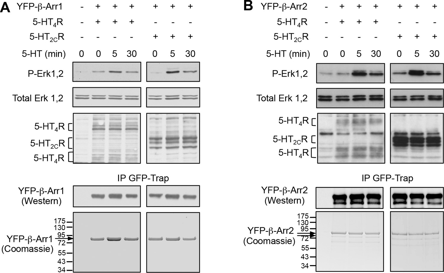
Purification of YFP-β-arrestin1 and YFP-β-arrestin2 co-expressed with 5-HT2C or 5-HT4 receptor in HEK-293 cells.
YFP-β-arrestin1 (A) and YFP-β-arrestin2 (B) co-expressed with Myc-tagged 5-HT2C or 5-HT4 receptor were immunoprecipitated using GFP Trap beads and detected by Western blotting using an anti-GFP antibody (10% of IP) and by colloidal Coomassie blue staining (90% of IP). Receptor expression and functionality were assessed by immunoblotting using an anti-Myc antibody, and by sequential immunoblotting with the antibody recognizing phospho-Thr202/Tyr204-Erk1/2 and total Erk1/2. Immunoblots and gels representative of four independent experiments are illustrated. Note that 5-HT4 receptor immunoreactivity was detected at molecular weights corresponding to receptor monomer and dimer.
Figure 1—figure supplement 2
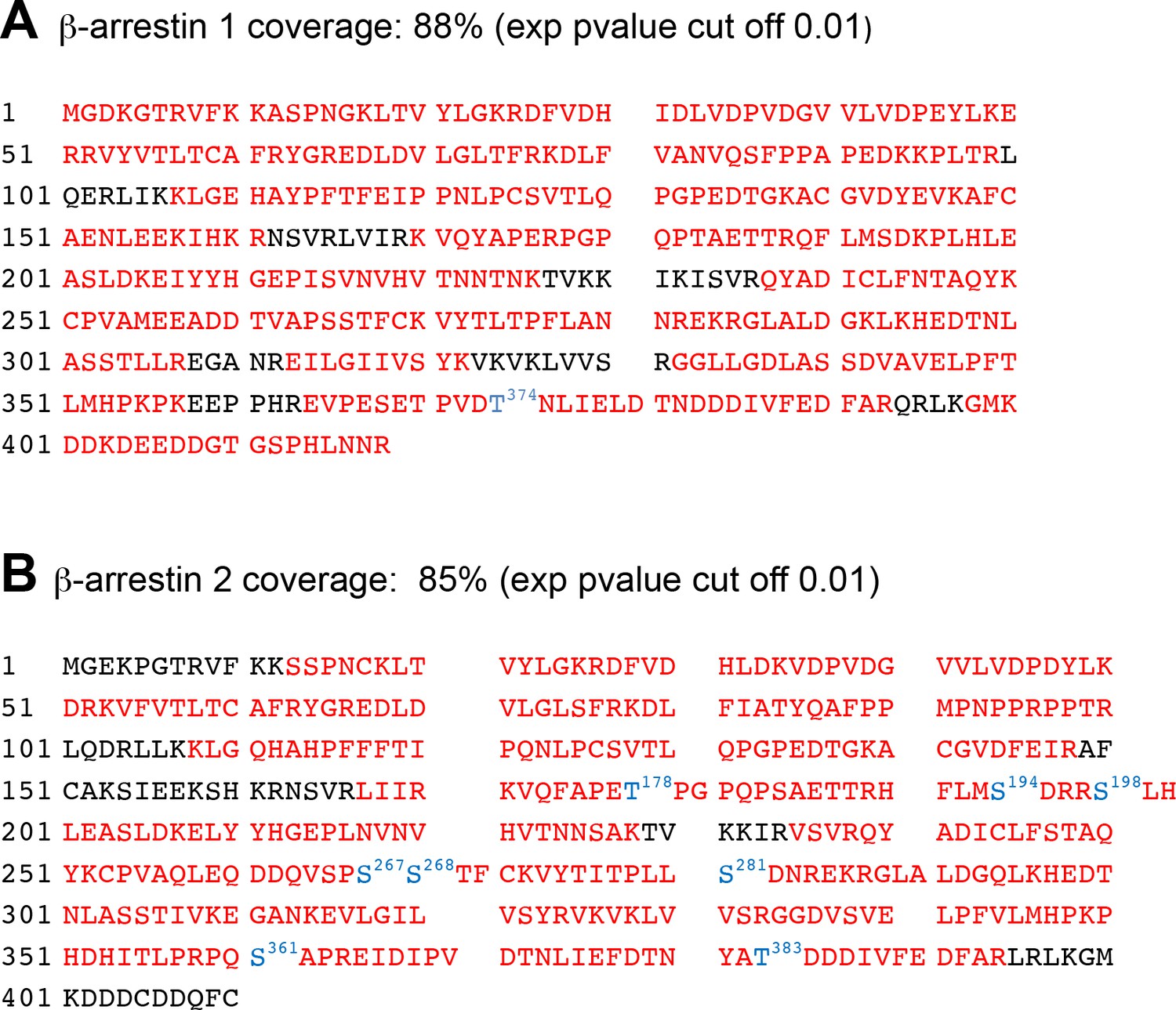
Sequence coverage of β-arrestin1 and β-arrestin2 obtained by LC-MS/MS.
The sequence covered by LC-MS/MS analysis is highlighted in red. Identified phosphorylated residues in β-arrestin1 (Thr374) and β-arrestin2 (Thr178, Ser194, Ser267/268, Ser281, Ser361 and Thr383) and their positions are highlighted in blue.
Figure 1—figure supplement 3
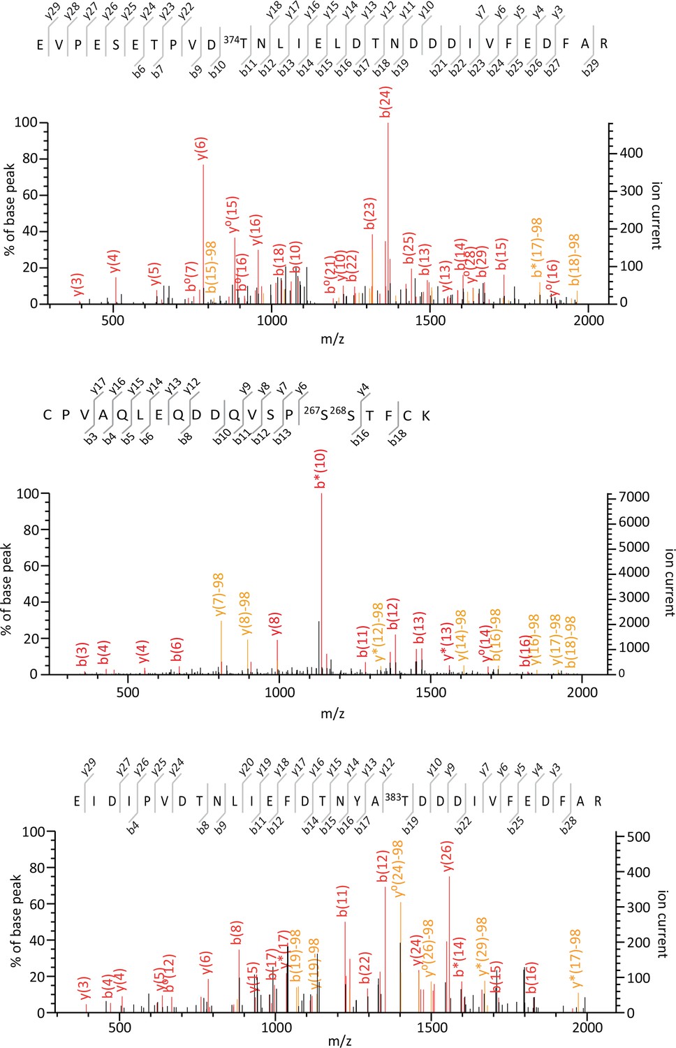
Tandem mass spectra of EVPESETPVDpT374NLIELDT NDDDIVFEDFAR, CPVAQLEQDDQVSPp(S267S268)TFCK and EIDIPVDTNLIEFD TNYApT383DDDIVFEDFAR phosphorylated peptides identified from YFP-tagged β-arrestin1 and β-arrestin2 transiently co-expressed with 5-HT2C receptor in HEK-293 cells and immunoprecipitated using the GFP Trap kit.
For each identified phosphorylated peptide, MS/MS spectra that yielded the highest Mascot score, matched b and y ions, peptide sequence and position of the phosphorylated residue in the full-length protein are illustrated.
Figure 1—figure supplement 4
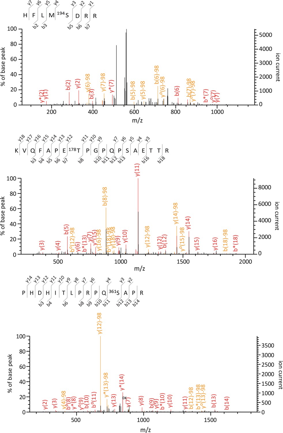
Tandem mass spectra of HFLMpS194DRR, KVQFAPE pT178PGPQPSAETTR and PHDHITLPRPQpS361APR phosphorylated peptides identified from YFP-tagged β-arrestin1 and β-arrestin2 transiently co-expressed with 5-HT2C receptor in HEK-293 cells and immunoprecipitated using the GFP Trap kit.
For each identified phosphorylated peptide, MS/MS spectra that yielded the highest Mascot score, matched b and y ions, peptide sequence and position of the phosphorylated residue in the full-length protein are illustrated.
Figure 1—figure supplement 5
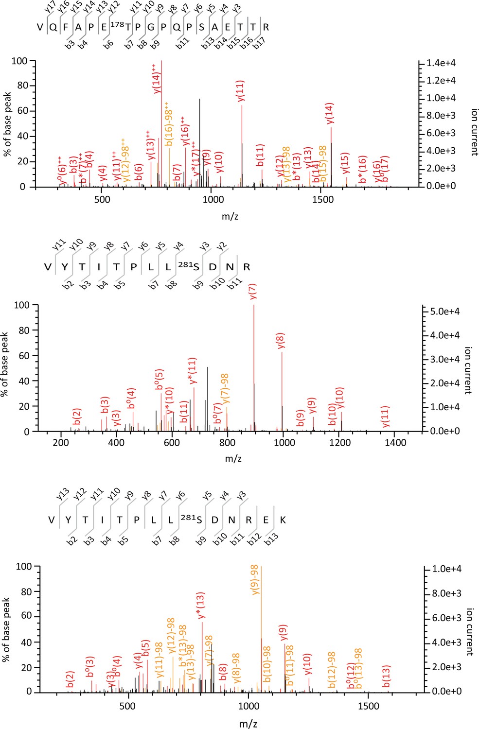
Tandem mass spectra of VQFAPEpT178PGPQPSAET TR, VYTITPLLpS281DNR, VYTITPLLpS281DNREK phosphorylated peptides identified from YFP-tagged β-arrestin1 and β-arrestin2 transiently co-expressed with 5-HT2C receptor in HEK-293 cells and immunoprecipitated using the GFP Trap kit.
For each identified phosphorylated peptide, MS/MS spectra that yielded the highest Mascot score, matched b and y ions, peptide sequence and position of the phosphorylated residue in the full-length protein are illustrated.
Figure 2 with 3 supplements
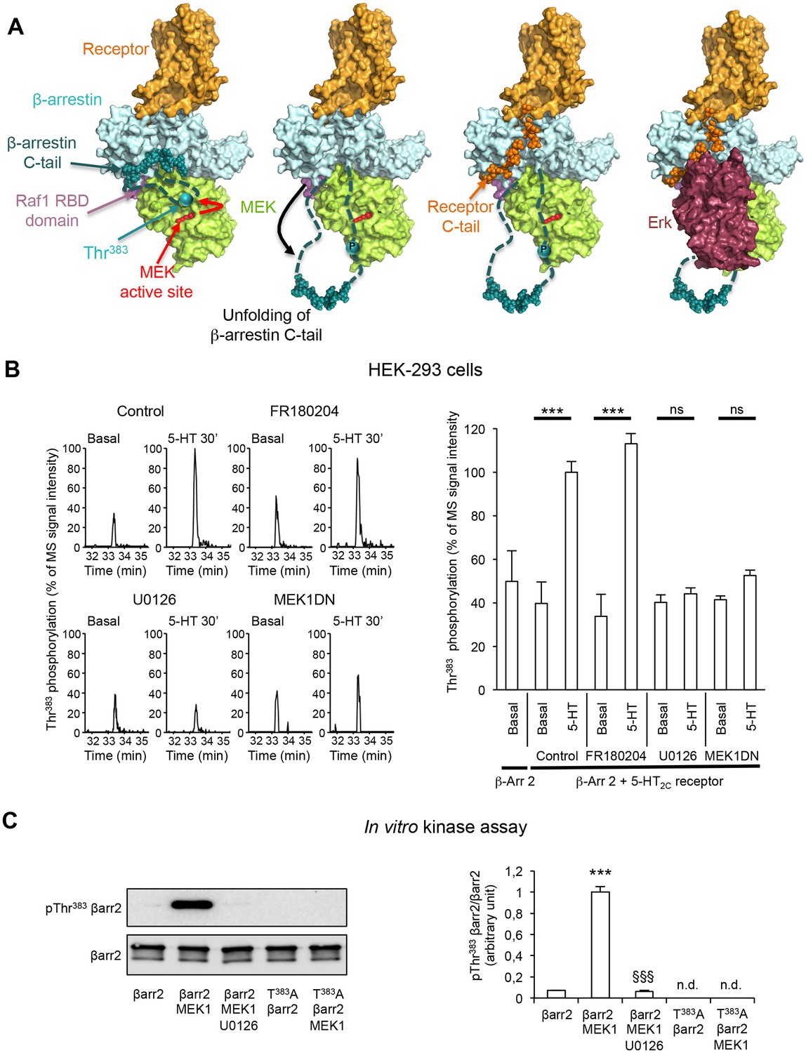
Role of MEK in the phosphorylation of β-arrestin2 at Thr383 elicited by 5-HT2C receptor stimulation.
(A) Mechanistic model of assembly of the 5-HT2C receptor/β-arrestin2/Erk module. Color code: receptor in orange, MEK in green, β-arrestin2 core in pale cyan and C-tail in cyan (the regions 351–384 and 394–419, which are not visible in 3D structure are represented by dashed lines, the region 385–393 is represented by spheres), Erk in dark red, Raf-1 RBD domain in pink. In this model, we hypothesize that Thr383 phosphorylation by MEK takes place within the assembled receptor/β-arrestin/Raf/MEK complex and results in a movement of β-arrestin2 unfolded 350–393 segment away from the first β-strand of β-arrestin, leaving space for further interaction with the receptor C-terminal domain (orange spheres) and recruitment of Erk, and its subsequent phosphorylation by MEK. For the clarity of the figure, the extremity of the β-arrestin C-tail is represented by spheres even in its unfolded state, although the real 3D structure is unknown. (B) Representative extracted ion chromatograms of the peptide in cells expressing 5-HT2C receptor, pretreated with either vehicle (control) or FR180204 (10 µM for 18 hr) or U0126 (5 µM for 30 min) or coexpressing MEK1 dominant-negative mutant (MEK1DN), and challenged with vehicle (Basal) or 5-HT (1 µM) for 30 min. The histogram represents the means ± SEM of the corresponding ion signal intensities (normalized to values in 5-HT-stimulated cells in Control condition) obtained in three independent experiments. One-way ANOVA: F(8,18) = 15.69, p<0.0001. ***p<0.001 vs. corresponding basal value. (C) YFP-tagged β-arrestin2 (wild-type or Thr383Ala mutant) purified from transfected HEK-293 cells was incubated with active MEK1 for 15 min at 37°C. When indicated, U0126 (5 µM) was included in the incubation medium. Thr383 phosphorylation was assessed by sequential immunoblotting with the antibody raised against phospho-Thr383 β-arrestin2 and the anti-β-arrestin2 antibody. Means ± SEM of results from four independent experiments are shown on the histogram. n.d.: not detectable. One-way ANOVA: F(2,9) = 352.2, p<0.0001. ***p<0.001 vs. immunoreactive signal in absence of MEK; §§§ p<0.001 vs. corresponding condition in absence of U0126.
-
Figure 2—source data 1
- https://doi.org/10.7554/eLife.23777.011
Figure 2—figure supplement 1
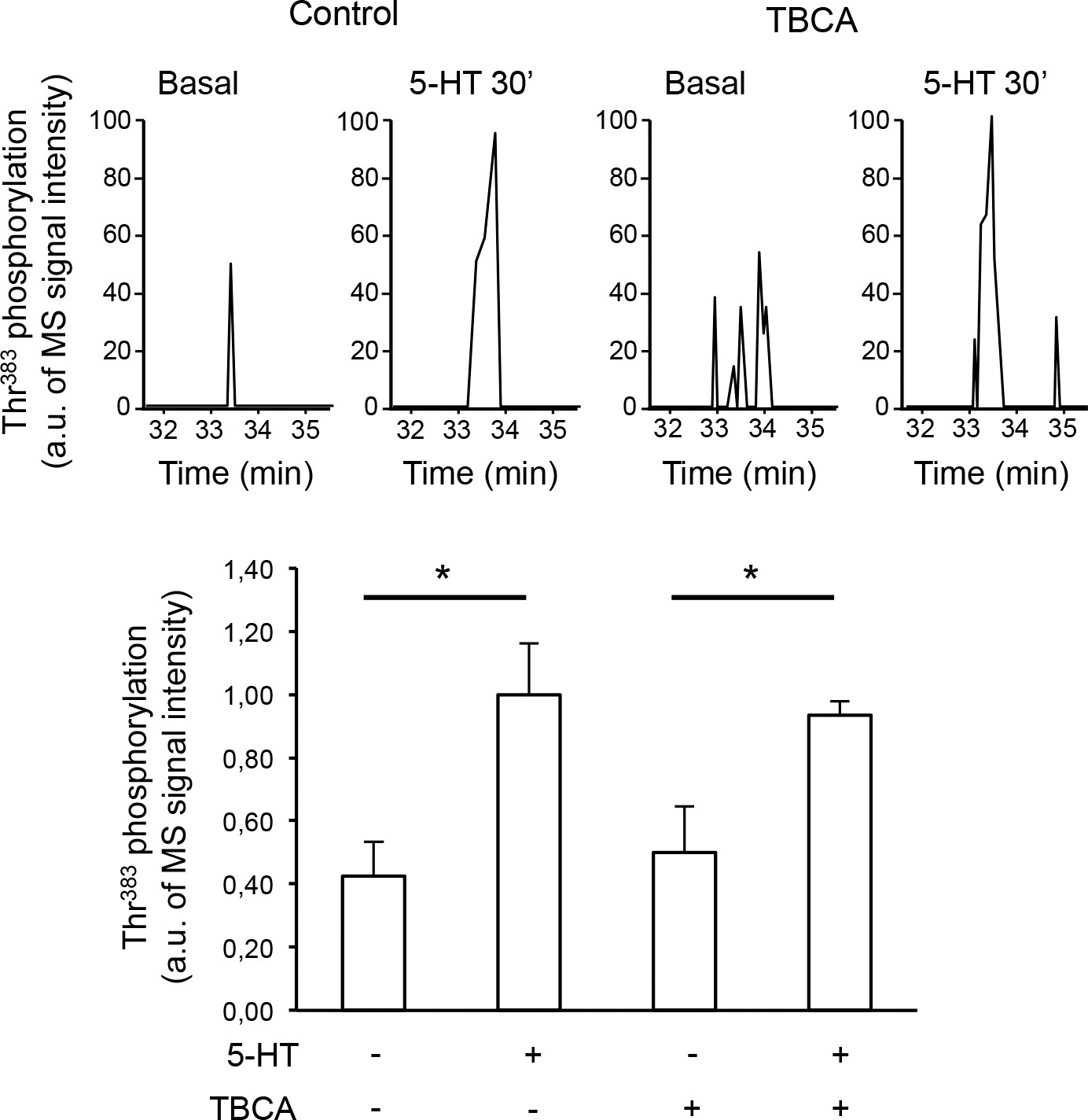
Thr383 phosphorylation is not mediated by casein kinase 2.
(A) Representative extracted ion chromatograms of the EIDIPVDTNLIEFDTNYAp383TDDDIVFEDFAR peptide from YFP-tagged β-arrestin2 in cells expressing 5-HT2C receptor and challenged with vehicle (Basal) or 1 µM 5-HT for 30 min in absence or presence of TBCA (1 µM, added 15 min before the onset of 5-HT treatment). Two other independent experiments performed on different sets of cultured cells yielded similar results. (B) The histogram represents the means ± SEM of ion signal intensities of the peptide (normalized to values in 5-HT-stimulated cells) obtained in the three experiments. One-way ANOVA F(3,8) = 6.762, p=0.013. *p<0.05 vs. corresponding vehicle.
-
Figure 2—figure supplement 1—source data 1
This file contains raw values used to build Figure 2—figure supplement 1.
- https://doi.org/10.7554/eLife.23777.013
Figure 2—figure supplement 2
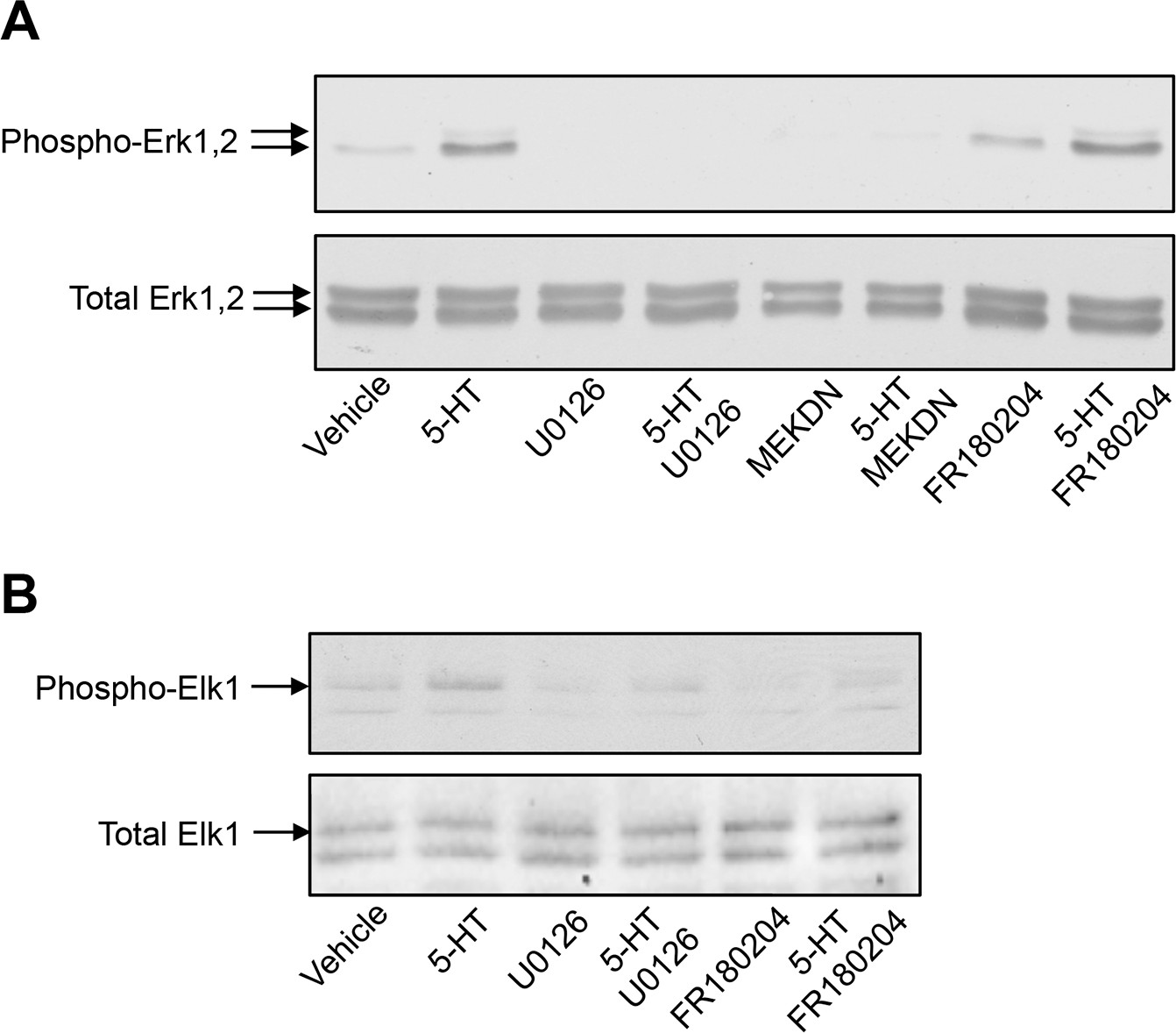
Impact of MEK and Erk1/2 pharmacological inhibitors and of co-expression of MEK dominant-negative mutant on 5-HT2C receptor-operated Erk1/2 and Elk1 phosphorylation.
(A) Cells expressing 5-HT2C receptor were pretreated with vehicle or U0126 (5 µM for 30 min) or FR180204 (10 µM for 18 hr) and then exposed to vehicle or 5-HT (1 µM) for 5 min. When indicated, a dominant-negative mutant of MEK (MEKDN) was co-expressed by 5-HT2C receptor. Erk1/2 activation was assessed by sequential immunoblotting with the antibody recognizing phospho-Thr202/Tyr204-Erk1/2 and total Erk1/2. Immunoblots representative of three independent experiments are illustrated. Note that FR180184 did not affect 5-HT2C receptor-operated Erk1/2 phosphorylation, indicating that it does not inhibit MEK. (B) The impact of MEK and Erk1/2 inhibitors on phosphorylation of the Erk1/2 substrate Elk1 was assessed by sequential immunoblotting with the antibody recognizing phospho-Elk1 and total Elk1. Immunoblots representative of two independent experiments are illustrated.
Figure 2—figure supplement 3
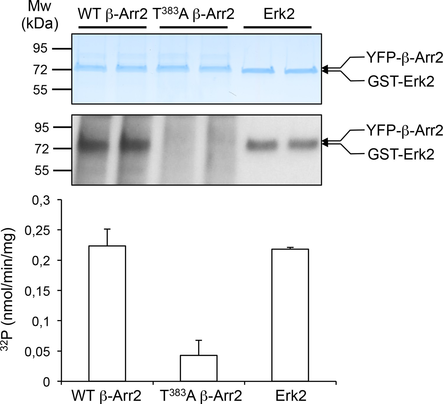
In vitro phosphorylation of β-arrestin2 versus Erk2 by MEK1.
YFP-tagged β-arrestin2 (wild-type or Thr383Ala mutant) purified from transfected HEK-293 cells or purified non-activated Erk2 (~1 µg each) were incubated with active MEK1 in presence of [γ-32P]-ATP (2 µCi/nmol) for 10 min at 37°C. Proteins were separated by SDS-PAGE and stained with Coomassie colloidal blue (top image) and 32P incorporation into the different substrates was monitored by autoradiography (bottom image). The data in the histogram, expressed in nmol/min/mg enzyme, represent the means ± SD of 32P incorporation into β-arrestin2 and Erk2 protein bands in the corresponding experiment after radioactive background subtraction for each lane.
-
Figure 2—figure supplement 3—source data 1
This file contains raw values used to build Figure 2—figure supplement 3.
- https://doi.org/10.7554/eLife.23777.016
Figure 3

Thr383 phosphorylation underlies β-arrestin2 conformational rearrangement elicited by 5-HT2C receptor stimulation.
(A, B) Translocation of wild type (WT), T383A and T383D Rluc-β-arrestin2 to Myc-5-HT2C-YFP (A) or Myc-5-HT4-YFP (B) receptors in cells treated with either vehicle (Basal) or 1 or 10 µM 5-HT, respectively, was measured by BRET. Data represent the mean ± SEM of values obtained in three independent experiments and were normalized to the BRET signals measured in 5-HT-stimulated cells expressing WT Rluc β-arrestin2. (C, D) Cell surface expression of receptors was measured in the same experimental condition by ELISA using anti-Myc antibody. Data are the mean ± SEM of values obtained in three independent experiments. They were normalized to total receptor expression level and are expressed in % of basal receptor level at the cell surface in cells expressing WT β-arrestin2. (E, F) Conformational arrangement of WT, T383A and T383D double brilliance Rluc8-β-arrestin2-RGFP elicited by 5-HT2C and 5-HT4 receptor stimulation by 5-HT (1 and 10 µM, respectively). Equivalent expression of each BRET sensor was verified by ELISA. Data represent the mean ± SEM of values obtained in three independent experiments and were normalized to the basal intra-molecular BRET signal in cells expressing WT Rluc8-β-arrestin2-RGFP. One-way ANOVA: A, F(5,12)=10.75, p=0.0004; B, F(5,12)=320.9, p<0.001; C, F(6,14)=10.82, p<0.0001; D, F(6,14)=48.52, p<0.0001; E, F(5,12)=5.136, p=0.0095; F, F(5,12)=6.436, p=0.004. *p<0.05, **p<0.01 ***p<0.001 vs. corresponding basal.
-
Figure 3—source data 1
This file contains raw values used to build Figure 3.
- https://doi.org/10.7554/eLife.23777.018
Figure 4 with 1 supplement
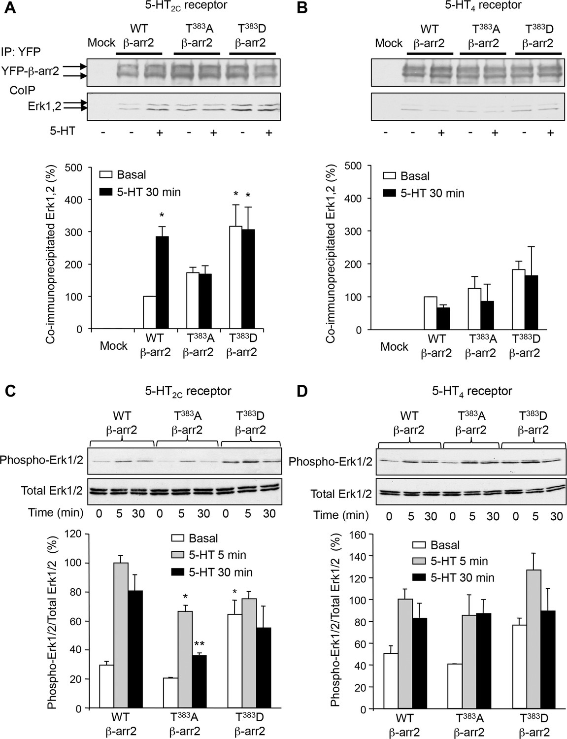
Phosphorylation of β-arrestin2 at Thr383 is a necessary step in Erk1/2 recruitment to β-arrestin2 and engagement of Erk signaling by 5-HT2C receptor.
(A, B) Recruitment of Erk1/2 to WT, T383A and T383D YFP-β-arrestin2 in cells expressing 5-HT2C or 5-HT4 receptor and exposed or not to 5-HT (1 and 10 µM, respectively) was assessed by co-immunoprecipitation. Immunoblots representative of three independent experiments are illustrated. The histograms represent the means ± SEM of Erk1/2 immunoreactive signals in immunoprecipitates, assessed by densitometric analysis, obtained in the three experiments. They were normalized to the amount of YFP-β-arrestin2 immunoprecipitates and expressed in % of basal level measured in cells expressing WT β-arrestin2. *p<0.05 vs. basal value in cells expressing WT β-arrestin2. (C, D) Erk1/2 activation in cells co-expressing 5-HT2C or 5-HT4 receptor and either WT, or T383A and or T383D YFP-β-arrestin2 and exposed or not to 5-HT (1 and 10 µM, respectively) for 5 or 30 min was assessed by sequential immunoblotting with the antibody recognizing phospho-Thr202/Tyr204-Erk1/2 and total Erk1/2. Immunoblots representative of three independent experiments are illustrated. The histograms represent the means ± SEM of values (normalized to the level of phosphorylated Erk1/2 in cells expressing WT β-arrestin2 and exposed to 5-HT for 5 min) obtained in the three experiments. One-way ANOVA: A, F(5,12)=4.305, p=0.00178; B, F(5,12)=0.977, p=0.47; C, F(8,18)=11.78, p<0001; D, F(8,18)=4.998, p=0.0022. *p<0.05, **p<0.01 vs. corresponding value in cells expressing WT β-arrestin2.
-
Figure 4—source data 1
This file contains raw values used to build Figure 4.
- https://doi.org/10.7554/eLife.23777.020
Figure 4—figure supplement 1
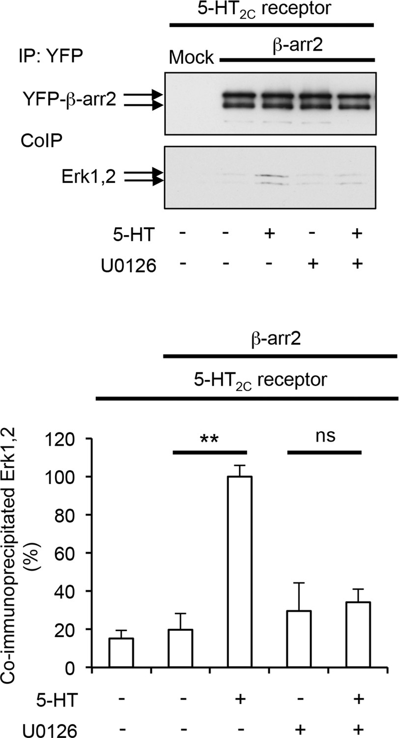
Role of MEK in Erk recruitment to β-arrestin2.
HEK-293 cells co-expressing YFP-β-arrestin2 and 5-HT2C receptor were treated for 30 min with vehicle or 1 µM 5-HT in absence or presence of 5 µM U0126 (added 15 min before the onset of 5-HT application). Recruitment of Erk1/2 to β-arrestin2 was assessed by co-immunoprecipitation. Immunoblots representative of three independent experiments are illustrated. The histogram represents the means ± SEM of Erk1/2 immunoreactive signals in immunoprecipitates normalized to the amount of YFP-β-arrestin2 immunoprecipitates, obtained in the three experiments. They were expressed in % of Erk immunoreactive signal measured in 5-HT-stimulated cells. One-way ANOVA: F(4,10) = 12,21, p=0.0007. **p<0.01 vs. vehicle.
-
Figure 4—figure supplement 1—source data 1
This file contains raw values used to build Figure 4—figure supplement 1.
- https://doi.org/10.7554/eLife.23777.022
Figure 5 with 1 supplement
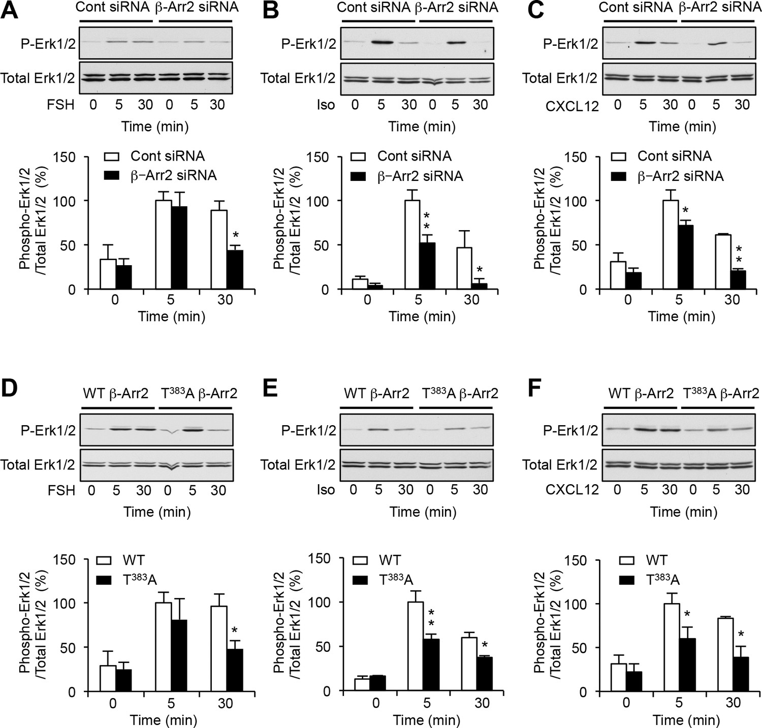
Phosphorylation of β-arrestin2 at Thr383 underlies β-arrestin-dependent engagement of Erk1/2 signaling by FSH, β-adrenergic and CXCR4 receptors.
Erk1/2 activation elicited by stimulation for the indicated times of transiently expressed FSH receptor (FSH, 3.3 nM), native β-adrenergic (isoproterenol 1 µM) and CXCR4 receptors (CXCL12, 10 nM) in cells transfected with control siRNA or β-arrestin2 siRNA (A–C) and in cells coexpressing WT or T383A β-arrestin2 (D–F). Erk1/2 activation was assessed by sequential immunoblotting with the antibody recognizing phospho-Thr202/Tyr204-Erk1/2 and total Erk1/2. Immunoblots representative of three independent experiments are illustrated. The histograms represent the means ± SEM of values (normalized to the level of phosphorylated Erk1/2 in cells exposed to agonist for 5 min) obtained in the three experiments. One-way ANOVA: A, F(5,12)=8.178, p=0.0014; B, F(5,12)=16.97, p<0.001; C, F(5,12)=20.22, p=<0.001; D, F(5,12)=7.710, p=0.0019; E, F(5,12)=29.76, p<0.0001; F(5,12)=8.695, p=0.0.0012. *p<0.05 and **p<0.01 vs. corresponding value in control siRNA transfected cells or cells expressing WT β-arrestin2.
-
Figure 5—source data 1
This file contains raw values used to build Figure 5.
- https://doi.org/10.7554/eLife.23777.024
Figure 5—figure supplement 1
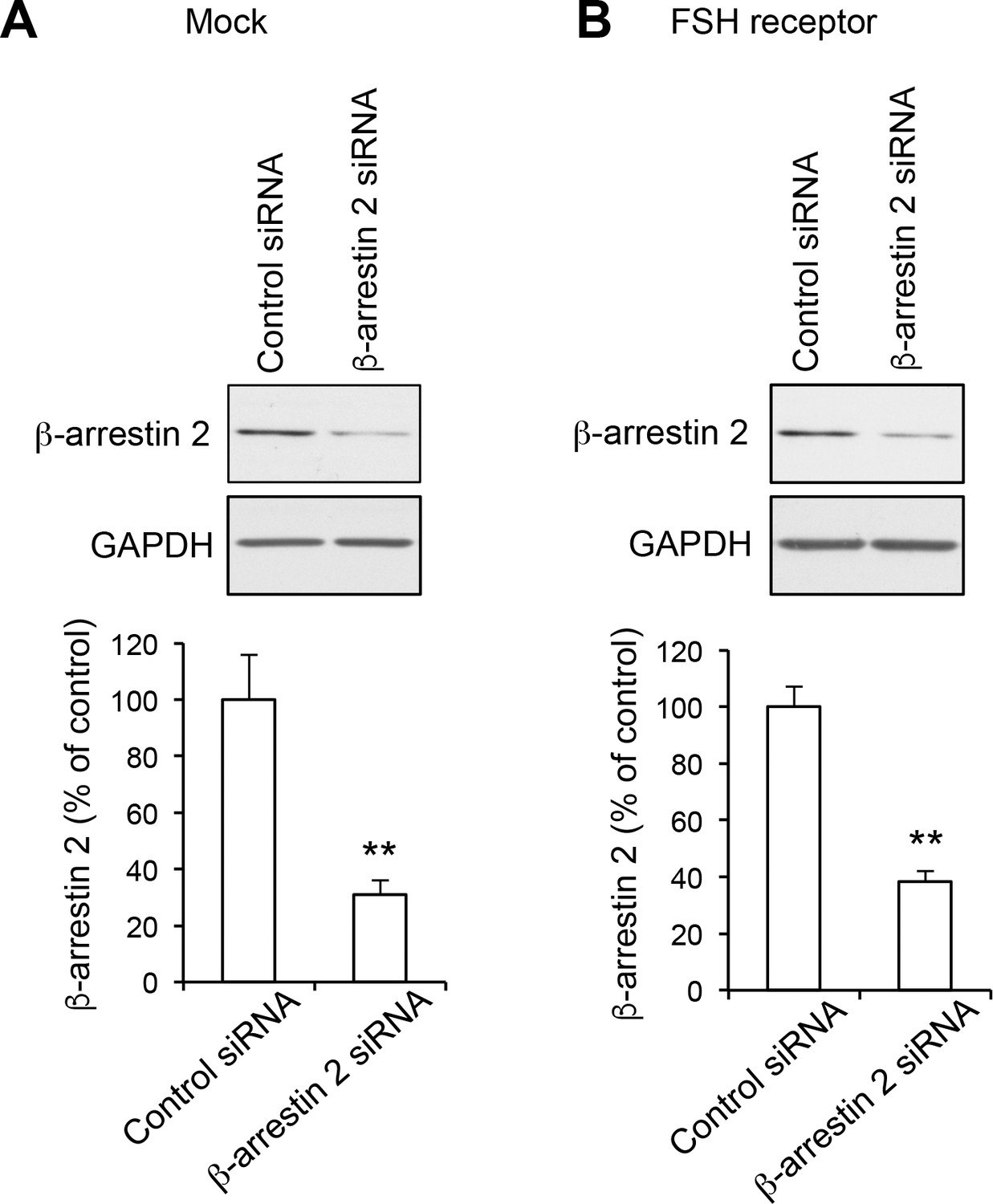
Down-regulation of β-arrestin2 protein expression in HEK-293 cells using siRNA.
Cells were transfected with either control or β-arrestin2 siRNA in absence (Mock, A) or presence (B) of the plasmid encoding FSH receptor. Cells were lysed 72 hr after transfection and expression of β-arrestin2 was measured by Western blotting. Equal loading was assessed by immunoblotting using a GAPDH antibody. Representative immunoblots of three independent experiments are illustrated. The histograms show the quantification of β-arrestin2 immunoreactivity in the corresponding experiments. Data, expressed in % of value measured in cells transfected with control siRNA, are means ± SEM of results obtained in three independent experiments. Unpaired t-test, A, p=0.008 vs. control siRNA; B, p=0.0017 vs. control siRNA.
-
Figure 5—figure supplement 1—source data 1
This file contains raw values used to build Figure 5—figure supplement 1.
- https://doi.org/10.7554/eLife.23777.026
Figure 6
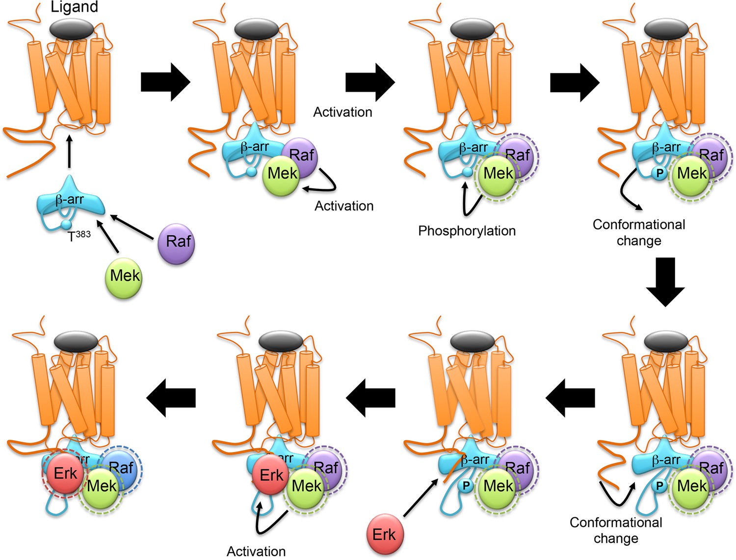
Schematic representation of the sequence of events proposed for β-arrestin-dependent Erk1/2 activation by GPCRs.
β-arrestin, Raf and MEK are recruited to agonist-stimulated receptor, resulting in MEK activation. Active MEK phosphorylates β-arrestin2 at Thr383. This induces a movement of the β-arrestin2 350–393 segment away from the first β-strand of β-arrestin, leaving space for its interaction with the C-terminal domain of the receptor. Erk then binds to the complex and is activated by MEK. The dash circles represent activated enzymes.
Download links
A two-part list of links to download the article, or parts of the article, in various formats.
Downloads (link to download the article as PDF)
Open citations (links to open the citations from this article in various online reference manager services)
Cite this article (links to download the citations from this article in formats compatible with various reference manager tools)
Phosphorylation of β-arrestin2 at Thr383 by MEK underlies β-arrestin-dependent activation of Erk1/2 by GPCRs
eLife 6:e23777.
https://doi.org/10.7554/eLife.23777


