A tRNA modification in Mycobacterium tuberculosis facilitates optimal intracellular growth
- Department of Immunology and Infectious Diseases Harvard T. H. Chan School of Public Health, United States
- Division of Infectious Diseases, Brigham and Women's Hospital, United States
- Department of Microbiology, Harvard Medical School, United States
- Howard Hughes Medical Institute, United States
Abstract
Diverse chemical modifications fine-tune the function and metabolism of tRNA. Although tRNA modification is universal in all kingdoms of life, profiles of modifications, their functions, and physiological roles have not been elucidated in most organisms including the human pathogen, Mycobacterium tuberculosis (Mtb), the causative agent of tuberculosis. To identify physiologically important modifications, we surveyed the tRNA of Mtb, using tRNA sequencing (tRNA-seq) and genome-mining. Homology searches identified 23 candidate tRNA modifying enzymes that are predicted to create 16 tRNA modifications across all tRNA species. Reverse transcription-derived error signatures in tRNA-seq predicted the sites and presence of nine modifications. Several chemical treatments prior to tRNA-seq expanded the number of predictable modifications. Deletion of Mtb genes encoding two modifying enzymes, TruB and MnmA, eliminated their respective tRNA modifications, validating the presence of modified sites in tRNA species. Furthermore, the absence of mnmA attenuated Mtb growth in macrophages, suggesting that MnmA-dependent tRNA uridine sulfation contributes to Mtb intracellular growth. Our results lay the foundation for unveiling the roles of tRNA modifications in Mtb pathogenesis and developing new therapeutics against tuberculosis.
eLife assessment
This is a valuable addition to the literature as it helps us understand the role of tRNA modifying enzymes in Mycobacterium tuberculosis. By knocking out one of the enzymes, the authors convincingly demonstrate the importance of tRNA-modifying enzymes for intra-host growth of tubercle bacteria. Some of the claims regarding modification as well as the role in virulence could be strengthened through further bioinformatics and phylogenetic analyses as well as experimental approaches. The work will be of interest to microbiologists.
https://doi.org/10.7554/eLife.87146.3.sa0Introduction
tRNA is an adaptor molecule that enables protein synthesis by converting the triplet genetic code in mRNA into amino acids. The fidelity of base pairing between mRNA codons and tRNA anticodons is monitored within ribosomes and is critical for properly incorporating the amino acids bound to the 3′ ends of tRNAs into growing polypeptides. For optimal translation, the abundances, and properties of tRNA isoacceptors are fine tuned by diverse mechanisms, including chemical modifications (Huang and Hopper, 2016; Björk and Hagervall, 2014; Shepherd and Ibba, 2015; Torrent et al., 2018). Dysregulation of tRNA abundance and/or structure leads to defective decoding and results in ribosome pausing and collisions, protein misfolding, stress responses and can have detrimental or lethal effects on the cell (Orellana et al., 2022; Suzuki, 2021; Liu et al., 2022; Goodarzi et al., 2016; Delaunay et al., 2022; Nedialkova and Leidel, 2015).
Chemical modifications of tRNA (tRNA modifications) are found in all kingdoms of life and fine-tune tRNA properties including mRNA decoding efficiency, recognition by aminoacyl-tRNA synthetases, half-life, and structural stability (Björk and Hagervall, 2014; Nedialkova and Leidel, 2015; Kimura and Waldor, 2019; Giegé and Eriani, 2023). Modifications are prevalent in the anticodon loop, particularly at the first letter of the anticodon. Modifications of the anticodon loop directly modulate codon recognition, whereas modifications in the tRNA body region primarily stabilize tRNA tertiary structure, protecting them from degradation in the cell. tRNA modifications are generated by dedicated site-specific enzymes referred to as tRNA modifying enzymes. tRNA modifications have been extensively characterized in a few model organisms (Björk and Hagervall, 2014; de Crécy-Lagard and Jaroch, 2021), but their profiles, regulation, and functions in non-model organisms, including bacterial pathogens, are understudied (de Crécy-Lagard and Jaroch, 2021).
tRNA sequencing (tRNA-seq) allows for the rapid and systematic prediction of many tRNA modification sites (Zhang et al., 2022; Zheng et al., 2015). We recently developed a comparative tRNA-seq protocol to profile tRNA modifications in organisms with uncharted tRNA modification profiles; in Vibrio cholerae, this approach led to the discovery of a new RNA modification and RNA editing process (Kimura et al., 2020). tRNA-seq enables rapid prediction of modified sites through detection of reverse transcription-derived signatures, such as nucleotide misincorporation and early termination, both of which occur more frequently at modified sites. Furthermore, several chemical treatments of tRNA can convert modifications that are not recognizable as reverse transcription-derived signatures into detectable signals, expanding the repertoire of modifications that can be distinguished by tRNA-seq (Motorin and Helm, 2019; Finet et al., 2022; Draycott et al., 2022; Dai et al., 2023).
Mycobacterium tuberculosis (Mtb), the agent of tuberculosis (TB), is a global pathogen that caused >10.5 million cases and over 1.5 million deaths worldwide in 2020 (W.H.O, 2021). Several studies have uncovered roles for non-Mtb mycobacterial tRNA modifications in stress responses, adaptation to environmental changes, and persister cell formation (Chionh et al., 2016). Mycobacterium bovis BCG, an organism closely related to Mtb, responds to hypoxia by reprogramming 40 ribonucleoside modifications in tRNA to facilitate translation of a subset of proteins that promote survival in hypoxic conditions (Chionh et al., 2016). To date, studies of the profiles and functions of Mtb tRNA modifications have been limited.
Here, we conducted tRNA-seq in Mtb. We assigned modifications to many of the reverse transcription-derived signatures identified, using information on the presence of homologs of known modifying enzymes in Mtb. Chemical treatments of tRNAs carried out prior to tRNA-seq increased the detectability of certain modifications. We constructed two Mtb deletion mutant strains, with deletions of mnmA and truB, and confirmed that the absence of these modification enzymes eliminated the predicted signals in tRNA-seq data. Furthermore, while deletion of mnmA in Mtb did not affect the pathogen’s growth in in vitro laboratory growth conditions, the mnmA knockout strain’s growth was attenuated in a macrophage infection model. Our findings suggest that tRNA modifications warrant further study as we unravel the complexity of Mtb infections, as they may serve as targets for new therapeutics.
Results
In silico prediction of Mtb tRNA modifying enzymes
To predict tRNA modifications in Mtb, we used Basic Local Alignment Search Tool (BLAST) (Altschul et al., 1990) to identify homologs of all tRNA modification enzymes registered in Modomics in the Mtb genome (Boccaletto et al., 2022; Supplementary file 1). With a stringent threshold (E-value <1 × 10⁻¹⁰), 31 Mtb genes homologous to genes encoding known RNA modification enzymes were identified (Supplementary file 2). Twenty-three of these genes are predicted to synthesize 16 tRNA modifications in Mtb (Supplementary files 2 and 3), including miaA and miaB for 2-methylthio-6-isopentenyl-adenosine (ms2i6A), tsaD, tsaB, tsaE, and sua5 for N6-threonylcarbamoyladenosine (t6A), mnmA for 2-thiouridine (s2U), truB, truA, rluA, and pus9 for pseudouridine (Ψ), trm2 for 5-methyluridine (m5U), trmD for 1-methylguanosine (m1G), trmI for 1-methyladenosine (m1A), trmL for 2′-O-methylcytidine (Cm) or 2′-O-methyluridine (Um), trmB for 7-methylguanosine (m7G), two trmH for 2′-O-methylguanosine (Gm), trmR for 5-methoxyuridine (mo5U), trcM for 5-methylcytidine (m5C), dusB for dihydrouridine (D), tadA for inosine (I), and tilS for lysidine (k2C). While eight additional genes met the threshold for homology to modification enzymes, they exhibited greater similarity to a Glutamyl-tRNA synthase; Rv2992c, ribosome associated GTPases (Der; Rv1713 and Era; Rv2364c), molybdopterin biosynthesis proteins (MoeW; Rv2338c and MoeB2; Rv3116), thiosulfate sulfur transferases (CysA; Rv3117 and SseA; Rv3283) and a riboflavin biosynthesis protein (RibG; Rv1409), and likely do not correspond to tRNA modification enzymes (Supplementary file 2).
We mined data from genome-wide Tn-seq and CRISPRi screens (DeJesus et al., 2017; Bosch et al., 2021) to assess the impacts of Mtb tRNA modifications on its growth. Five genes encoding Mtb tRNA modifying enzymes, trmD, tilS, tadA, tsaC2, and miaA, were reported to be essential for Mtb growth in both Tn-seq (DeJesus et al., 2017) and CRISPRi screens (Bosch et al., 2021; Supplementary file 3), suggesting that the modifications they produce are critical for tRNA functions. Indeed, E. coli trmD, tilS, tadA, and tsaC2 are also essential for growth and critical for codon decoding, aminoacylation, and reading frame maintenance (Masuda et al., 2022; Soma et al., 2003; Wolf et al., 2002; El Yacoubi et al., 2009). The Mtb modifications synthesized by these enzymes likely have similar impacts on tRNA functions. Unexpectedly, one modifying enzyme, MiaA, is non-essential in E. coli grown in nutrient-rich medium, but apparently essential in Mtb, suggesting that i6A, the modification introduced by MiaA, may have more profound roles in Mtb translation than in E. coli.
Several of the putative Mtb tRNA modifying enzymes are conserved across all three domains of life (e.g., TruB and TsaD) (Figure 1). By contrast, some enzymes were limited to bacterial species closely related to Mtb, possibly suggesting their species-specific physiological roles, including pathogenesis. For example, TrmI (Rv2118c) homologs are widely present in Archaea and Eukaryotes, but are sparsely distributed in bacteria, where they are primarily limited to Actinomycetia, including Mtb, and several thermophilic bacterial species (e.g., Thermus thermophilus). TrmI synthesizes m1A at position 58 in eukaryotes (Anderson et al., 1998) and at position 57/58 in archaea (Guelorget et al., 2010); indeed, TrmI in Mtb has been proven to generate m1A at position 58 (Varshney et al., 2004). Pus9 (Rv3300c) has many homologs exhibiting weak similarity across eukaryotes and bacteria. However, some species, including Pseudomonas aeruginosa, Acinetobacter baumannii, Neisseria gonorrhoeae, and several species of Actinomycetia, encode proteins showing strong homology to Mtb Pus9, suggesting that this set of pseudouridylases have a distinctive property such as substrate specificity.
Figure 1

Phylogenetic distribution of Mtb tRNA modifying enzyme homologs.
Heatmap of log10 E-values from BLAST search results. BLAST searches were conducted against 120 manually picked organisms using Mtb tRNA modifying enzymes as queries. When one organism has multiple hits, the lowest log10(Eval) values among hits are shown. iTol (Letunic and Bork, 2021) was used to depict the results.
-
Figure 1—source data 1
log10 E-values from BLAST searching for the homologs of Mtb tRNA modifying enzymes in 120 organisms.
- https://cdn.elifesciences.org/articles/87146/elife-87146-fig1-data1-v1.xlsx
tRNA modifying enzymes predicted in Mtb were observed in mycobacteria species, including Mycolicibacterium smegmatis and Mycobacteroides abscessus (Figure 1), suggesting that tRNA modification patterns are similar among mycobacterium species.
Profiling Mtb tRNA modification sites by tRNA-seq
To begin profiling detectable Mtb tRNA modifications, we sequenced tRNAs isolated from wild-type Mtb strain H37Rv grown in 7H9 medium. In this protocol, tRNAs are first reversed transcribed to cDNA (Kimura et al., 2020). During cDNA synthesis, chemical modifications on tRNA nucleotides disrupt Watson–Crick base pairing and increase the frequency of reverse transcriptase errors, leading to incorporation of the incorrect nucleotide or early termination of cDNA synthesis (Kellner et al., 2010). These reverse transcription-derived ‘signatures’ typically correspond to modified sites (Zhang et al., 2022; Zheng et al., 2015) and are depicted in the heatmap in Figure 2.
Figure 2 with 1 supplement see all

Heatmap of misincorporation and early termination frequency in sequencing of tRNAs from wild-ype Mtb.
Heatmaps show misincorporation (A) and termination (B) frequencies at all positions across tRNAs (read 5′ to 3′). Predicted modifications are labeled based on similarity to known modifications in other organisms and the presence of the tRNA modifying enzyme homologs (Supplementary file 3). The positions with more than 10% misincorporation in Mtb but not in E. coli are depicted in white in A. Representative data of two independent experiments with similar results are shown. (C) M. tuberculosis tRNA modifications predicted in this study. Schematic tRNA secondary structure with sites of modifications identified either by the presence of modifying enzymes and/or tRNA sequencing (tRNA-seq). Modifications and tRNA species that are not observed in E. coli are shown in red. Modifications and positions that are predicted by both RT-derived signature and the presence of the homologs of tRNA modifying enzymes are shown in yellow (without chemical treatment) and green (with chemical treatment), whereas modifications that are only predicted by the presence of the homologs are shown in light blue. Genes reported to be essential in Mtb are shown in bold.
-
Figure 2—source data 1
Misincorporation and early termination frequencies in sequencing of tRNAs from wild-type Mtb.
- https://cdn.elifesciences.org/articles/87146/elife-87146-fig2-data1-v1.xlsx
Comparison of the reverse transcription-derived signatures observed in Mtb to E. coli, where tRNA modifications have been well characterized (Kimura et al., 2020) enables the prediction of the presence of common modifications, including ms2i6A, m1G, I, and k2C (Figure 2 and Figure 2—figure supplement 1). These predictions are strongly supported by the set of tRNA modification enzymes identified in the Mtb genome (Figure 1 and Supplementary file 3), including miaA and miaB (ms2i6A), trmD (m1G), tadA (I), and tilS (k2C). Some tRNA modifications were observed in Mtb but are not present in E. coli. In other actinobacteria, A58 and A59 are likely modified to m1A (Schwartz et al., 2018), and since trmI, the methylase that generates this modification is present in Mtb (Varshney et al., 2004), most Mtb tRNAs likely contain this modification as well. Nucleoside variation in the sequence of tRNA genes can account for some of the variations in modified sites between E. coli and Mtb. For example, in Mtb, termination signatures derived from G at position 37 were detected in tRNA-Arg2, -Gln1, and -Gln2, whereas this position in these tRNAs in E. coli are modified A, such as m2A and m6A, which are silent in tRNA-seq. Since m1G induces strong termination during reverse transcription, these positions are likely modified to m1G, as observed in the Bacillus subtilis tRNA-Arg2 gene position 37 G (Jühling et al., 2009).
tRNA samples were also treated with several chemical treatments prior to sequencing, to expand the set of tRNA modifications detectable by tRNA-seq. These treatments included iodoacetamide (IAA), for detection of sulfur modifications, 1-cyclohexyl-(2-morpholinoethyl) carbodiimide (CMC) for detection of Ψ, and alkali for detection of D and m7G. The chemical treatment protocols were first carried out with E. coli, to validate the methods.
IAA is a thiol-reactive compound that covalently attaches carboxyamidomethyl to thiolated uridines via nucleophilic substitution (Herzog et al., 2017) and modified s4U is detected as C instead of U. In IAA-treated samples, positions 8 and 9, corresponding to s4U in many tRNA species, had high misincorporation frequencies, confirming that IAA treatment modifies s4U, leading to elevated misincorporation (Figure 3—figure supplement 1). Furthermore, we observed higher misincorporation and termination signals at the positions corresponding to other sulfur modification, s2C, s2U and their derivatives, such as position 32 in tRNA-Arg3, -Arg5, -Ser3, and Arg4, and 34 in tRNA-Glu, -Gln1, and -Lys (Figure 3 and Figure 3—figure supplement 1), revealing that IAA treatment facilitates the detection of not only s4U but also additional sulfur modifications, which are weakly detected without the IAA treatment.
Figure 3 with 1 supplement see all
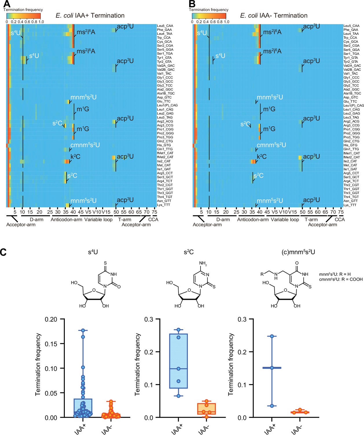
Iodoacetamide (IAA) treatment promotes detection of sulfur modifications on tRNAs by enhancing termination signals.
Heatmaps of the termination signals of E. coli tRNAs treated with (A) or without (B) IAA. Known modification sites, including sulfur modifications (s4U, s2C, s2U in white) are shown. (C) Termination frequency at s4U (n = 48), s2C (n = 5), and s2U (n = 3) sites of tRNAs treated with or without IAA. The experiment was performed once.
-
Figure 3—source data 1
Termination frequencies of E. coli tRNAs treated with or without iodoacetamide (IAA).
- https://cdn.elifesciences.org/articles/87146/elife-87146-fig3-data1-v1.xlsx
Next, we applied IAA treatment to Mtb tRNA-seq. IAA treatment increased termination signals from position 34 in tRNA-Glu1, -Gln1, and -Lys, which contain s2U derivatives in E. coli (Figure 4A). The 2-thiouridine modification is carried out by MnmA in E. coli (Kambampati and Lauhon, 2003), and a homolog, Rv3024c, of this enzyme was identified in the Mtb genome (Supplementary file 3; Kapopoulou et al., 2011). We used double-stranded DNA-based recombineering to delete Rv3024c in Mtb, yielding strain MtbΔmnmA (van Kessel and Hatfull, 2007). Sequencing of tRNA isolated from MtbΔmnmA with prior IAA treatment showed reduced termination signals from position 34 in tRNA-Glu1, -Gln1, and -Lys in treated samples, indicating that Rv3024c plays a critical role in the modification responsible for increased termination frequency derived from position 34 in these tRNAs (Figure 4B, C). Together, these observations strongly suggest that Rv3024c encodes an MnmA-like enzyme that sulfurates position 34 uridines in three Mtb tRNA isoacceptors.
Figure 4

Heatmap and plot of early termination frequency from sequencing of tRNAs from wild-type and MtbΔmnmA with and without RNA alkylation.
Heatmap of early termination frequencies across tRNA molecules and positions for wild-type (WT) (A) and MtbΔmnmA (B). Sulfur modification is shown in white. (C) Plot of termination frequencies at position 37 in WT Mtb and MtbΔmnmA for lysine_UUU, glutamate_UUG, and glutamine_UUC isoacceptors (n = 3). IAA: iodoacetamide. The experiment was performed once.
-
Figure 4—source data 1
Termination frequencies from sequencing of tRNAs from wild-type and MtbΔmnmA with and without RNA alkylation.
- https://cdn.elifesciences.org/articles/87146/elife-87146-fig4-data1-v1.xlsx
As shown previously (Carlile et al., 2014), CMC treatment increased both misincorporation and termination signatures at a subset of Ψs in E. coli tRNAs (Figure 5, and Figure 5—figure supplement 1 and Figure 5—figure supplement 2). In addition, CMC-treated samples showed increased frequencies of both misincorporation and termination at positions 16, 17, 20, and 20A corresponding to D, and m7G at position 46. Both modifications are known to undergo base elimination in mild alkali conditions (Marchand, 2021). Since these signals were also observed in the reaction condition in which CMC was not added, these signals are likely attributable to the alkali treatment that is common to both conditions.
Figure 5 with 4 supplements see all
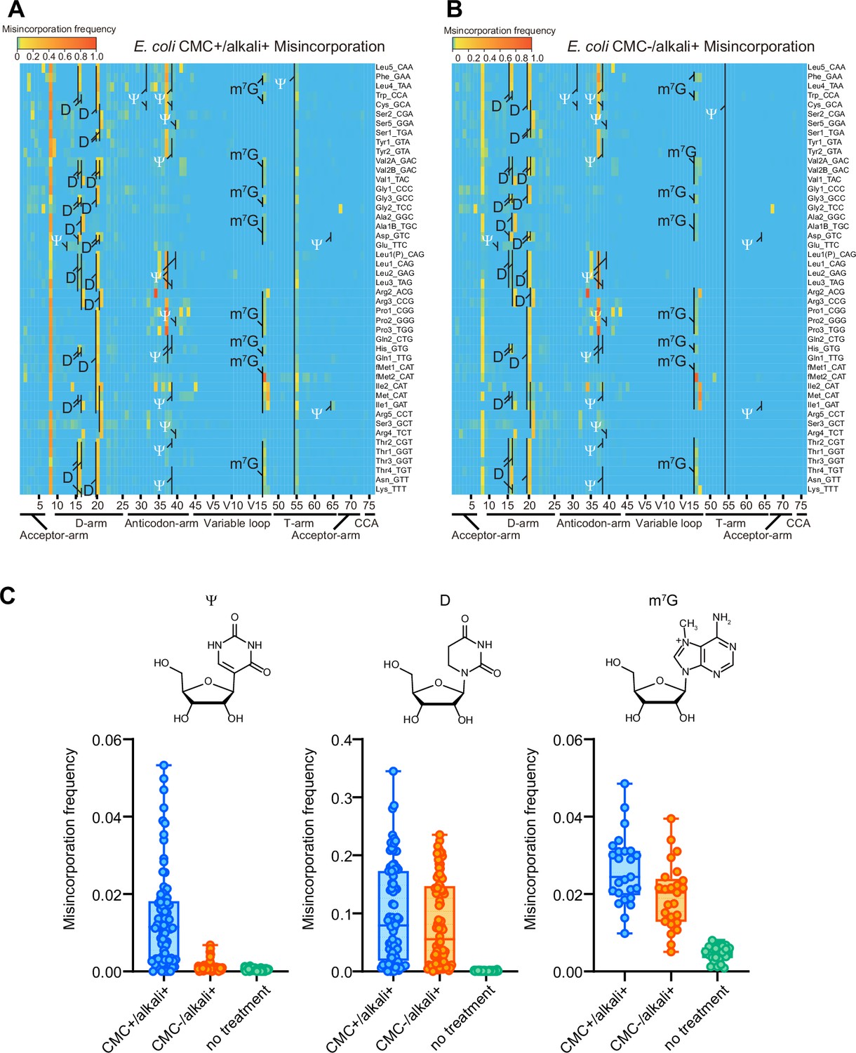
1-Cyclohexyl-(2-morpholinoethyl) carbodiimide (CMC) and alkali treatment facilitate detection of ψ, D, and m7G modifications in E. coli.
Heatmaps of the misincorporation signals of E. coli tRNAs treated with (A) or without (B) CMC. In both conditions, tRNAs are incubated in alkali condition. Known Ψ, D, and m7G sites are shown. Ψ is shown in white. (C) Misincorporation frequency at known Ψ (n = 80), D (n = 76), and m7G (n = 25) sites of tRNAs treated with CMC+/alkali+ or CMC−/alkali+, and tRNAs without treatment. The experiment was performed once.
-
Figure 5—source data 1
Misincorporation frequencies from sequencing of E. coli tRNAs treated with or without 1-cyclohexyl-(2-morpholinoethyl) carbodiimide (CMC).
- https://cdn.elifesciences.org/articles/87146/elife-87146-fig5-data1-v1.xlsx
Reverse transcription-derived signatures derived from alkali-treated D showed a distinctive pattern. With this treatment, when two Ds are at consecutive positions, for example, D16 and D17, termination signals were elevated at the following position, that is, position 18 (Figure 5—figure supplement 1, Figure 5—figure supplement 3, and Figure 5—figure supplement 4). Furthermore, alkali treatment also led to higher misincorporation frequencies at singlet Ds (Figure 5, Figure 5—figure supplement 3, and Figure 5—figure supplement 4). Thus, termination and misincorporation signatures enabled the prediction of known E. coli tRNA sites modified to D.
CMC/alkali treatment facilitated the identification of additional modifications in Mtb tRNAs. Regardless of CMC treatment, alkali-treated samples showed increased misincorporation and termination frequencies derived from U located at positions 16, 17, 20, and 20A (Figure 6 and Figure 6—figure supplement 1). As observed in E. coli, termination signals at positions 18 and 21 likely correspond to consecutive Ds at positions 16 and 17, and 20 and 20A, respectively. Mtb Rv0823c is a homolog of dihydrouridylase DusB (Supplementary file 3), which likely accounts for the synthesis of D at these positions. Furthermore, alkali treatment increased the misincorporation frequencies at G at position 46 (Figure 6—figure supplement 2). Since Rv0208c is a homolog of TrmB, which synthesizes m7G at position 46 in E. coli, multiple Mtb tRNA species likely contain m7G at position 46 (Figure 6 and Figure 6—figure supplement 2).
Figure 6 with 3 supplements see all

Heatmap of early termination frequency from sequencing of tRNAs isolated from wild-type (WT) and MtbΔtruB following 1-cyclohexyl-(2-morpholinoethyl) carbodiimide (CMC) treatment.
Heatmap of early termination frequencies across tRNA molecules and positions for WT (left) and MtbΔtruB (right). Termination signals derived from position 55 are shown in white. The experiment was performed once.
-
Figure 6—source data 1
Termination frequencies across tRNA molecules and positions for wild-type (WT) and MtbΔtruB.
- https://cdn.elifesciences.org/articles/87146/elife-87146-fig6-data1-v1.xlsx
CMC treatment also increased the termination frequency at several sites. Termination signatures derived from position 55, which is exclusively uridine in all tRNA species, increased in most tRNA species, suggesting that Mtb tRNAs contain pseudouridines at this position (Jühling et al., 2009). Rv2793c is an Mtb homolog of E. coli TruB and deletion of Rv2793c reduced the termination frequencies at this position in tRNAs isolated from MtbΔtruB (Figure 6 and Figure 6-figure supplement 3; van Kessel and Hatfull, 2007). Together, these observations suggest that Rv2793c encodes a TruB-like enzyme that modifies position 55 uridines to Ψ across tRNA species. Furthermore, the presence of a TruA homolog in Mtb (Rv3455c) suggests that in multiple tRNA species U at positions 38–40 can be modified to Ψ. Indeed, the termination signatures derived from positions 38 and 39 increased depending on CMC treatment, strongly suggesting that these positions are modified to Ψ (Figure 6 and Figure 6—figure supplement 3).
In total, among 16 tRNA Mtb tRNA modifications predicted by the presence of tRNA modifying enzymes (Figure 1 and Supplementary file 3), 9 species of modifications were detected based on reverse-transcription-derived signatures (Figure 2C).
Growth of MtbΔmnmA is attenuated in a macrophage infection model
To address whether Mtb tRNA modifications impact the pathogen’s growth in the host environment, we used the MtbTnDB transposon insertion sequencing (Tn-seq) database (Zhang et al., 2013; Jinich et al., 2021) to determine if transposon insertions in genes encoding tRNA modifying enzymes have been associated with in vivo growth defects. Transposon insertions in mnmA were reported to attenuate Mtb growth in mice infected with a library of Mtb transposon mutants, suggesting that mnmA facilitates Mtb growth in vivo. We found that the growth of WT and MtbΔmnmA were similar in 7H9 medium (Figure 7), suggesting the absence of s2U modification at position 34 does not impair Mtb growth in culture. In contrast, the MtbΔmnmA mutant was significantly impaired for growth in a macrophage infection model (Jinich et al., 2021; Figure 7). Defective growth of the MtbΔmnmA mutant was also observed in macrophages treated with all-trans retinoic acid (ATRA), which promotes macrophage control of Mtb infection (Babunovic et al., 2022). These observations strongly suggest that modification of U to s2U by MnmA facilitates Mtb growth in macrophages.
Figure 7
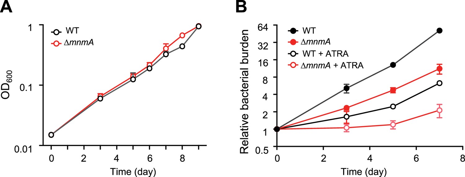
MtbΔmnmA is attenuated in a macrophage infection model.
(A) Wild-type and MtbΔmnmA do not display growth differences in 7H9 medium. (B) Auto-luminescent wild-type and ΔmnmA Mtb strains were diluted to a multiplicity of infection of 2 bacteria per mouse bone marrow-derived macrophage with or without all-trans retinoic acid (ATRA). Survival was measured by luminescence and normalized to luminescence at time 0. Average values from three independent cultures (n=3) are shown with standard deviations.
-
Figure 7—source data 1
Mtb growth curve.
- https://cdn.elifesciences.org/articles/87146/elife-87146-fig7-data1-v1.xlsx
Discussion
Here, we profiled Mtb tRNA modifications with tRNA-seq to provide the first maps of the tRNA modification landscape in this global pathogen. In total, nine modifications, including six modifications without chemical treatment, were identified based on reverse transcription-derived signatures. CMC/alkali treatment and IAA treatment further identified Ψ and m7G and sulfur modifications, respectively. Although we did not chemically validate the modifications predicted by tRNA-seq with mass spectrometry, the identification of Mtb homologs of tRNA modifying enzymes strongly bolsters the RT-signature-based predictions. Furthermore, the deletion of truB and mnmA genes in Mtb eliminated the respective modification signatures of pseudouridine and s2U, validating that the enzymes encoded by these genes synthesize these modifications. Finally, the growth defect of the ΔmnmA strain within macrophages but not in a nutrient-rich medium suggests that s2U tRNA modification facilitates Mtb adaptation to the host intracellular environment.
IAA treatment was developed for detecting s4U modification in pulse-chase experiments to measure RNA turnover (Herzog et al., 2017). We found that this treatment enhances the RT signatures of additional sulfur modifications as well, including s2C and s2U, indicating that IAA should have general utility in tRNA-seq-based profiling of sulfur modifications, including in studies tracking changes in tRNA sulfuration (Edwards et al., 2022; Laxman et al., 2013).
Multiple modifications do not cause reverse transcriptase errors. Although tRNA-seq provides a simple and rapid method for profiling the landscape of modifications in all tRNA species, this approach does not enable comprehensive identification of modifications. Here, several modifications, including t6A, m5U, mo5U, m5C, Cm, Um, and Gm, predicted by the presence of Mtb homolog of tRNA modifying enzymes, such as TsaC2, TsaB, TsaD, TsaE, Trm2, TrcM, TrmR, TrmL, and TrmH, were not detected in tRNA-seq. Additionally, CMC treatment may not determine all Ψ positions, thus, the target of Rv3300 and Rv1540 remain unclear. Since these genes are similar to E. coli rluA, which also targets rRNA, these genes may target rRNAs instead of tRNAs. Mapping and further analysis of these modifications will require different approaches such as tRNA purification and RNA mass spectrometric analysis.
Several strong RT signatures were detected in Mtb tRNAs but not in E. coli (Figure 2A), including G45 in tRNA-Gly2. Most of these signals are strict misincorporations of C or T at A or G position, respectively. Since modifications at a purine position, for example, m1G and m1A, generally cause random misincorporation of the other three nucleosides, these Mtb-specific misincorporation signatures are not likely to be derived from modification. Further mass spectrometric analysis of purified tRNAs will be necessary to assess whether these signals are indeed derived from Mtb-specific modifications; such Mtb-specific modifications could be related to Mtb pathogenesis and/or survival in the host.
The role of s2U in Mtb appears to be unusual. s2U is a universally conserved modification observed in all three domains of life at the wobble position in the anticodon. This modification enhances the stacking of s2U with U35 to stabilize the anticodon structure, facilitating codon–anticodon interactions. s2U is also recognized by multiple aminoacyl-tRNA synthetases for efficient amino acylation (Giegé and Eriani, 2023). Although elimination of this sulfur modification causes severe growth phenotypes in most organisms (Kambampati and Lauhon, 2003; Dewez et al., 2008), unexpectedly, the deletion of mnmA in Mtb did not attenuate growth of the pathogen in vitro, suggesting that the Mtb requirement for s2U modification differs from other organisms. The marked growth retardation of ΔmnmA strain within macrophages indicates the specific requirement of this modification within host cells. This modification may be necessary for maintaining general translation efficiency inside host cells and/or facilitate the expression of specific genes that are necessary for survival within macrophages. In fact, lack of mnmA is reported to sensitize E. coli to oxidative stress, raising the possibility that s2U modification promotes Mtb growth under oxidative stress elicited by the host.
Differential codon usage between house-keeping genes and virulence genes could contribute to the differential growth phenotypes observed in vitro and in vivo in the ΔmnmA mutant. Among multiple codons encoding Glu, Gln, and Lys, s2U modification dependent codon usage might be preferentially distributed in genes associated with intracellular growth. For example, Mtb has two tRNA isoacceptors, tRNA-Glu1(s2UUC) and tRNA-Glu2(CUC), to decipher two Glu codons, GAA and GAG. According to the wobble paring rule, GAA is only decoded by tRNA-Glu1(s2UUC), whereas GAG is decoded by both tRNA-Glu1(s2UUC) and tRNA-Glu2(CUC); that is, GAG can be deciphered by an s2U-independent tRNA. Thus, genes required for intracellular growth might be enriched with GAA, an s2U-dependent codon. Similar codon usage differences could be present in Gln and Lys codons deciphered by s2U-containing tRNAs.
In most organisms, s2U is further modified into derivatives containing an additional chemical moiety at position 5 (Björk and Hagervall, 2014; Karlsborn et al., 2014; Asano et al., 2018). However, Mtb does not contain apparent homologs of the tRNA modifying enzymes that introduce the additional modifications to s2U. Thus, Mtb may contain s2U or s2U derivatives synthesized by other types of enzymes. Additional analyses to elucidate the structures of modified s2U in Mtb are warranted.
Another unexpected finding is the presence of a TrmR homolog in Mtb. TrmR in B. subtilis is a methylase that converts a 5-hydroxyuridine (ho5U) into a 5-methoxyuridine (mo5U) at the tRNA wobble position, suggesting that Mtb possesses mo5U. However, Mtb does not encode an apparent homolog for the enzyme that mediates the RNA hydroxylation reaction that yields ho5U, the substrate for TrmR. Although we cannot rule out the possibility that Mtb TrmR methylates a substrate other than RNA, it is possible that Mtb utilizes an unknown hydroxylation pathway for synthesizing ho5U.
Our findings serve as a valuable starting point for the research community to continue characterizing the physiological roles and mechanisms of Mtb tRNA modifications. Since tRNA-seq offers an efficient and scalable platform for surveying changes in tRNA modifications, this approach will be valuable across growth conditions, and may be extended to growth inside host cells. Finally, further studies elucidating the mechanisms by which tRNA modifications facilitate Mtb growth in host cells should be valuable for designing new therapeutics for tuberculosis.
Materials and methods
Key resources table
| Reagent type (species) or resource | Designation | Source or reference | Identifiers | Additional information |
|---|---|---|---|---|
| Gene (Mycobacterium tuberculosis) | mnmA | NA | Uniprot: Rv3024c; Refseq: NP_217540.1 | |
| Gene (Mycobacterium tuberculosis) | truB | NA | Uniprot: Rv2793c; Refseq: NP_217309.1 | |
| Strain, strain background (Mycobacterium tuberculosis) | H37Rv | PMID:9634230 | ||
| Genetic reagent (Mycobacterium tuberculosis) | MtbΔmnmA::zeo | This paper | Mtb H37Rv strain lacking mnmA | |
| Genetic reagent (Mycobacterium tuberculosis) | MtbΔtruB::zeo | This paper | Mtb H37Rv strain lacking truB | |
| Recombinant DNA reagent | pNit-RecET SacBR | NA | For homologous recombination | |
| Chemical compound | Iodoacetamide (IAA) | Sigma | ||
| Chemical compound | 1-Cyclohexyl-(2-morpholinoethyl) carbodiimide (CMC) | Sigma |
Bacterial strains and growth conditions
Request a detailed protocolMtb strains were grown from frozen stocks into Middlebrook 7H9 medium supplemented with 0.2% glycerol, 0.05% Tween-80, and ADC (5 g/l bovine serum albumin, 2 g/l dextrose, 3 μg/ml catalase). Cultures were incubated at 37°C. Strains were grown to mid-log phase for all experiments (OD600 0.4–0.6). Growth was measured on a BioTek plate reader for in vitro growth by measuring OD600 every 24 hr.
Bacterial strain construction
Supplementary file 4 depicts the strains, plasmids, primers, and recombinant DNA used for this study. Plasmids were built by restriction digest of a parental vector and inserts were prepared by Gibson assembly (Gibson et al., 2009). Plasmids were isolated from E. coli and confirmed via Sanger sequencing carried out by Genewiz, LLC (Massachusetts, USA).
Deletion mutants
Request a detailed protocolThe knockout strain Mtb∆mnmA::zeo (zeocin) was built using double-stranded recombineering in the parental Mtb strain H37Rv. A linear dsDNA fragment was constructed using stitch polymerase chain reaction (PCR) with the primers listed in Supplementary file 4 which consisted of a 500-bp region upstream of mnmA (Rv3024c), 500-bp downstream region, and a lox-zeo-lox fragment. This cassette was transformed into an H37Rv recombineering strain as described (Murphy et al., 2015) and plated on 7H10 + zeocin plates.
Homology search
Request a detailed protocolLocal BLAST was performed to search for Mtb homologs of tRNA modifying enzymes. First, the uniport IDs of tRNA modifying enzymes were obtained from Modomics (Boccaletto et al., 2022), and 12 proteins were manually added to the list (Cho et al., 2023; Kimura et al., 2020; Sakai et al., 2019; Kimura et al., 2022; Takakura et al., 2019; Sakai et al., 2016), including Q47319/TapT, P24188/TrhO, P76403/TrhP, O32034/TrhP1, O32035/TrhP2, P36566/CmoM, and Q87K36/TrcP, O34614/MnmM, Q8N5C7/DTWD1, Q8NBA8/DTWD2, O32036/TrmR, Q9KV41/AcpA. Uniprot ID provides a fasta file of tRNA modifying enzymes from the Uniprot database (UniProt, 2023). A blast database file was generated by ‘makeblastdb’ script using a fasta file of Mtb proteins (H37Rv strain) retrieved from NCBI. Then, the homologs of tRNA modifying enzymes were searched against the Mtb protein database using the fasta file of tRNA modifying enzymes as a query. Output format is defined by the following script: -outfmt ‘7 qacc sacc stitle score qcovs evalue pident’ -evalue 1e-10. The output file was modified by excel (Supplementary file 4).
Phylogenetic analysis of Mtb tRNA modifying enzyme homologs
Request a detailed protocolLocal BLAST was conducted to search for homologs of Mtb tRNA modifying enzymes in 120 manually picked organisms across three domains of life. A custom database was generated by combining the fasta files of organisms’ proteins retrieved from NCBI. Homologs of 18 Mtb tRNA modifying enzymes were searched against the custom protein database using local blast. Log10 E-values were visualized by iTol (Letunic and Bork, 2021) with a phylogenetic tree generated by phyloT (PhyloT, 2015).
tRNA sequencing
Extraction of total RNA
Request a detailed protocolStrains were grown to mid-log phase with the appropriate antibiotics and inducing agents described above. RNA was collected at the same OD600 for each strain (between 0.4 and .6). Cells were left on ice for 20 min, then pelleted by centrifuging at 4000 rpm for 10 min at 4°C. Pellets were resuspended in 0.5–1 ml of TriZol (Life Technologies) and lysed using a BeadBug microtube homogenizer (Millipore Sigma). 200 μl of chloroform was added to each tube, after which samples obtained from Mtb strains were removed from biosafety level 3 precautions. Samples were centrifuged at 15,000 rpm for 15 min at 4°C and the aqueous layer was collected into a fresh tube. To the original tube, 250 μl of sodium acetate buffer (300 mM sodium acetate pH 5.2 and 10 mM ethylenediaminetetraacetic acid (EDTA) pH 8.0) was added, and samples were vortexed at 4°C for 5 min then centrifuged at 15,000 × g for 15 min at 4°C. The aqueous layer was added to the fresh sample-containing tubes. 400 μl chloroform was added, and tubes were briefly vortexed and then centrifuged at 15,000 rpm for 1 min at 4°C. The aqueous phase was collected into a fresh tube and RNA recovered by ethanol precipitation. RNA pellets were resuspended in 10 mM sodium acetate pH 5.2 and stored at −80°C until processed for sequencing. Total RNA samples were alkali treated prior to tRNA extraction to deacylate all tRNAs (1 hr at 37°C in 100 mM Tris–HCl pH 9.0).
Isolation of tRNA fraction
Request a detailed protocol1–2 μg of total RNA was run on a 10% TBE-UREA gel (Thermo Fisher Scientific) at 250 V for 1 hr. Gels were stained with SYBR Gold (Thermo Fisher Scientific), and tRNA was excised. Excised gels containing tRNA fractions were mashed in RNAse-free tubes, and 300 μl elution buffer (300 mM NaOAc pH 5.5, 1 mM EDTA pH 8.0, 0.10% sodium dodecyl sulfate) was added to each tube. Samples were shaken on a thermoshaker (Eppendorf) for 1–4 hr at 37°C and supernatant was collected using an Ultrafree filter column (Millipore Sigma). tRNA was recovered by isopropanol precipitation.
tRNA dephosphorylation
Request a detailed protocoltRNA was dephosphorylated using QuickCIP (New England BioLabs) according to manufacturer instructions, and tRNA was collected by phenol–chloroform extraction followed by isopropanol precipitation.
IAA treatment
Request a detailed protocolIAA treatment was performed as described (Herzog et al., 2017). Briefly, 500 ng of total RNA is combined with 10 mM of IAA, 50 mM NaPO4 pH 8.0, and 50% dimethyl sulfoxide (DMSO) in a final volume of 50 μl. Reactions were incubated at 50°C for 15 min and quenched with dithiothreitol (DTT).
CMC treatment
Request a detailed protocolCMC treatment was carried out as described in ref Briefly, 2.5 μg tRNA fraction in 0.5 μl was mixed with 15 μl CMC-BEU buffer with or without CMC (0.34 M or 0 M CMC, 7 M urea, 4 mM EDTA, and 50 mM bicine pH 7.9) and incubated at 37°C for 20 min. Adding 100 μl CMC stop solution (0.3 M NaOAc pH 5.2 and 100 mM EDTA) quenched the reaction. RNA was desalted with PD-10 desalting column (Cytiva) and recovered by ethanol precipitation. RNA was dissolved in 40 μl of 50 mM sodium carbonate buffer (pH 10.4) and incubated at 37°C for 4 hr, followed by ethanol precipitation.
Adapter ligation
Request a detailed protocol0.5 μL RNase inhibitor was added to 3.5 μl dephosphorylated tRNA (200–250 ng tRNA) and samples were boiled at 80°C for 2 min. Boiled tRNA was mixed with 12 μl PEG buffer mix (10 μl 50% PEG8000, 2 μl 10× buffer B0216S; New England Biolabs). 3 μl of 5′ adenylated linkers (Supplementary file 4) were added (33 pmol/μl) along with 1 μl T4 RNA ligase 2 truncated (New England BioLabs) and incubated at 25°C for 2.5 hr. Samples were recovered by isopropanol precipitation and run on a 10% TBE-Urea PAGE gel for 40 min at 250 V. Ligated products were recovered by gel excision as described above.
Reverse transcription
Request a detailed protocolIdentical quantities of samples with different adapter sequences were pooled for reverse transcription for a total of 200–250 ng tRNA. Reverse transcription was performed by combining 2.1 μl dephosphorylated tRNA with 100 mM Tris–HCl pH 7.5, 0.5 mM EDTA, 1.25 μM RT primer (Supplementary file 4), 450 mM NaCl, 5 mM MgCl2, 5 mM DTT, 500 nM TGIRT (InGex), and 15% PEG8000 in a final volume of 9 μl. Samples were incubated at 25°C for 30 min, after which 1 μl 10 mM dNTPs (New England BioLabs) were added and reactions incubated at 60°C for 1 hr. 1.15 μl NaOH was added, and samples were boiled for 15 min and run on a 10% TBE Urea PAGE gel at 250 V for 1 hr. Reverse transcription products were excised from gels and cDNA recovered by isopropanol precipitation. Linear single-stranded cDNA was circularized using CircLigase II (Lucigen) in accordance with manufacturer instructions.
PCR of tRNA libraries
Request a detailed protocolPCR reactions were set up using HF Phusion according to the manufacturer’s instructions using a universal reverse primer (Supplementary file 4) and a different index primer for each pool of samples. PCR reactions were aliquoted into 4 tubes and collected after 6, 8, 10, and 12 cycles. Samples were run on a Native TBE PAGE gel (Thermo Fisher Scientific) at 180 V for 50 min, and amplified products were cut from the same cycle for each sequencing run. Samples were recovered by gel excision and isopropanol precipitation.
Sequencing
Request a detailed protocolSequencing was performed on a MiSeq instrument (Illumina) using 150 bp single end reads with a version 3, 150 cycle kit.
Analysis
3′ linker sequences and two nucleotides at the 5′ end were trimmed using cutadapt and fastx-trimmer. Bowtie v1.2.2 was used with default settings to map reads to reference Mtb tRNA sequences retrieved from Mycobrowser (Kapopoulou et al., 2011; Supplementary file 5). Mpileup files were generated using samtools (samtools mpileup -I -A --ff 4 -x -B -q 0 -d 10000000). For analysis of termination frequencies, 5′ end termini of mapped reads were piled up using bedtools genomecov (option, -d -5 -ibam). The number of 5′ termini at each tRNA position was divided by the total number of mapped termini at that position plus all upstream (5′) positions.
Macrophage infection
Request a detailed protocolAuto-luminescent wild-type and ΔmnmA Mtb strains grown to the same OD600 were pelleted by centrifugation and prepared in RPMI media by soft spinning as described (Saito et al., 2017). Briefly, cells were washed, pelleted, resuspended, and centrifuged at 121 × g, with the top half of the centrifuged supernatant used. Suspensions were diluted to a multiplicity of infection of 2 bacteria per mouse bone marrow-derived macrophage by determining the OD600. Macrophages were infected for 6 hr, followed by a phosphate-buffered saline wash and addition of RPMI with or without ATRA. ATRA promotes macrophage control of Mtb infection (Babunovic et al., 2022) and was used to assess strain survival in an increasingly restricted macrophage environment. Survival was measured by luminescence in a BioTek plate reader and normalized to luminescence reads at time 0.
Data availability
The sequencing data reported in this paper have been deposited in the NCBI Gene expression omnibus https://www.ncbi.nlm.nih.gov/geo/ (accession code, GSE240200).
-
NCBI Gene Expression OmnibusID GSE240200. Profiling tRNA modifications in Mycobacterium tuberculosis.
References
-
Basic local alignment search toolJournal of Molecular Biology 215:403–410.https://doi.org/10.1016/S0022-2836(05)80360-2
-
MODOMICS: a database of RNA modification pathways. 2021 updateNucleic Acids Research 50:D231–D235.https://doi.org/10.1093/nar/gkab1083
-
tRNA-mediated codon-biased translation in mycobacterial hypoxic persistenceNature Communications 7:13302.https://doi.org/10.1038/ncomms13302
-
Identification of a novel 5-aminomethyl-2-thiouridine methyltransferase in tRNA modificationNucleic Acids Research 51:1971–1983.https://doi.org/10.1093/nar/gkad048
-
Functions of Bacterial tRNA Modifications: from ubiquity to diversityTrends in Microbiology 29:41–53.https://doi.org/10.1016/j.tim.2020.06.010
-
Sulfur availability impacts accumulation of the 2-Thiouridine tRNA modification in Bacillus subtilisJournal of Bacteriology 204:e0000922.https://doi.org/10.1128/jb.00009-22
-
The universal YrdC/Sua5 family is required for the formation of threonylcarbamoyladenosine in tRNANucleic Acids Research 37:2894–2909.https://doi.org/10.1093/nar/gkp152
-
The tRNA identity landscape for aminoacylation and beyondNucleic Acids Research 51:1528–1570.https://doi.org/10.1093/nar/gkad007
-
Thiol-linked alkylation of RNA to assess expression dynamicsNature Methods 14:1198–1204.https://doi.org/10.1038/nmeth.4435
-
tRNAdb 2009: compilation of tRNA sequences and tRNA genesNucleic Acids Research 37:D159–D162.https://doi.org/10.1093/nar/gkn772
-
Comparative tRNA sequencing and RNA mass spectrometry for surveying tRNA modificationsNature Chemical Biology 16:964–972.https://doi.org/10.1038/s41589-020-0558-1
-
Interactive Tree Of Life (iTOL) v5: an online tool for phylogenetic tree display and annotationNucleic Acids Research 49:W293–W296.https://doi.org/10.1093/nar/gkab301
-
Mycobacterial recombineeringMethods in Molecular Biology 1285:177–199.https://doi.org/10.1007/978-1-4939-2450-9_10
-
tRNA dysregulation and diseaseNature Reviews. Genetics 23:651–664.https://doi.org/10.1038/s41576-022-00501-9
-
The expanding world of tRNA modifications and their disease relevanceNature Reviews. Molecular Cell Biology 22:375–392.https://doi.org/10.1038/s41580-021-00342-0
-
Biogenesis and functions of aminocarboxypropyluridine in tRNANature Communications 10:5542.https://doi.org/10.1038/s41467-019-13525-3
-
UniProt: the universal protein knowledgebase in 2023Nucleic Acids Research 51:D523–D531.https://doi.org/10.1093/nar/gkac1052
-
Recombineering in Mycobacterium tuberculosisNature Methods 4:147–152.https://doi.org/10.1038/nmeth996
-
SoftwareGlobal tuberculosis reportWorld Health Organisation.
-
tadA, an essential tRNA-specific adenosine deaminase from Escherichia coliThe EMBO Journal 21:3841–3851.https://doi.org/10.1093/emboj/cdf362
-
Efficient and quantitative high-throughput tRNA sequencingNature Methods 12:835–837.https://doi.org/10.1038/nmeth.3478
Article and author information
Author details
Funding
National Institute of Allergy and Infectious Diseases (R01AI-042347)
- Matthew K Waldor
National Institute of Allergy and Infectious Diseases (P01AI-095208)
- Eric J Rubin
Harvard Medical School (Dean's Innovation Award)
- Eric J Rubin
Howard Hughes Medical Institute (MKW)
- Matthew K Waldor
The funders had no role in study design, data collection, and interpretation, or the decision to submit the work for publication.
Acknowledgements
We appreciate all the members of Waldor lab for fruitful discussion and comments on the manuscript. We also thank Gregory Babunovic for his valuable assistance setting up the macrophage infections used here. This work is supported by NIH/NIAID grants to MKW (R01AI-042347) and EJR (P01AI-095208), a Dean’s Innovation Award from Harvard Medical School to EJR and the Howard Hughes Medical Institute (HHMI) to MKW.
Version history
- Preprint posted: February 20, 2023 (view preprint)
- Sent for peer review: February 27, 2023
- Preprint posted: April 12, 2023 (view preprint)
- Preprint posted: August 8, 2023 (view preprint)
- Version of Record published: September 27, 2023 (version 1)
Cite all versions
You can cite all versions using the DOI https://doi.org/10.7554/eLife.87146. This DOI represents all versions, and will always resolve to the latest one.
Copyright
© 2023, Tomasi, Kimura et al.
This article is distributed under the terms of the Creative Commons Attribution License, which permits unrestricted use and redistribution provided that the original author and source are credited.
Metrics
-
- 1,819
- views
-
- 111
- downloads
-
- 3
- citations
Views, downloads and citations are aggregated across all versions of this paper published by eLife.
Download links
A two-part list of links to download the article, or parts of the article, in various formats.
Downloads (link to download the article as PDF)
Open citations (links to open the citations from this article in various online reference manager services)
Cite this article (links to download the citations from this article in formats compatible with various reference manager tools)
A tRNA modification in Mycobacterium tuberculosis facilitates optimal intracellular growth
eLife 12:RP87146.
https://doi.org/10.7554/eLife.87146.3
Further reading
-
- Biochemistry and Chemical Biology
- Cell Biology
Mediator of ERBB2-driven Cell Motility 1 (MEMO1) is an evolutionary conserved protein implicated in many biological processes; however, its primary molecular function remains unknown. Importantly, MEMO1 is overexpressed in many types of cancer and was shown to modulate breast cancer metastasis through altered cell motility. To better understand the function of MEMO1 in cancer cells, we analyzed genetic interactions of MEMO1 using gene essentiality data from 1028 cancer cell lines and found multiple iron-related genes exhibiting genetic relationships with MEMO1. We experimentally confirmed several interactions between MEMO1 and iron-related proteins in living cells, most notably, transferrin receptor 2 (TFR2), mitoferrin-2 (SLC25A28), and the global iron response regulator IRP1 (ACO1). These interactions indicate that cells with high MEMO1 expression levels are hypersensitive to the disruptions in iron distribution. Our data also indicate that MEMO1 is involved in ferroptosis and is linked to iron supply to mitochondria. We have found that purified MEMO1 binds iron with high affinity under redox conditions mimicking intracellular environment and solved MEMO1 structures in complex with iron and copper. Our work reveals that the iron coordination mode in MEMO1 is very similar to that of iron-containing extradiol dioxygenases, which also display a similar structural fold. We conclude that MEMO1 is an iron-binding protein that modulates iron homeostasis in cancer cells.
-
- Biochemistry and Chemical Biology
- Structural Biology and Molecular Biophysics
NADPH oxidases (NOX) are transmembrane proteins, widely spread in eukaryotes and prokaryotes, that produce reactive oxygen species (ROS). Eukaryotes use the ROS products for innate immune defense and signaling in critical (patho)physiological processes. Despite the recent structures of human NOX isoforms, the activation of electron transfer remains incompletely understood. SpNOX, a homolog from Streptococcus pneumoniae, can serves as a robust model for exploring electron transfers in the NOX family thanks to its constitutive activity. Crystal structures of SpNOX full-length and dehydrogenase (DH) domain constructs are revealed here. The isolated DH domain acts as a flavin reductase, and both constructs use either NADPH or NADH as substrate. Our findings suggest that hydride transfer from NAD(P)H to FAD is the rate-limiting step in electron transfer. We identify significance of F397 in nicotinamide access to flavin isoalloxazine and confirm flavin binding contributions from both DH and Transmembrane (TM) domains. Comparison with related enzymes suggests that distal access to heme may influence the final electron acceptor, while the relative position of DH and TM does not necessarily correlate with activity, contrary to previous suggestions. It rather suggests requirement of an internal rearrangement, within the DH domain, to switch from a resting to an active state. Thus, SpNOX appears to be a good model of active NOX2, which allows us to propose an explanation for NOX2’s requirement for activation.


